Review
. 2010 Apr;11(4):252-63.
doi: 10.1038/nrn2804. Epub 2010 Feb 24.

Mapping the face in the somatosensory brainstem

Affiliations
Review

Mapping the face in the somatosensory brainstem

Reha S Erzurumlu et al. Nat Rev Neurosci. 2010 Apr.

Abstract

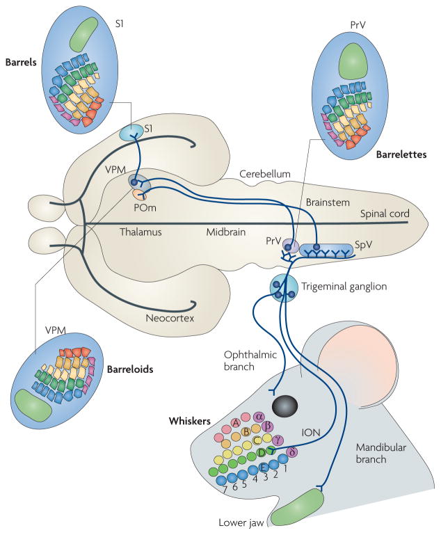
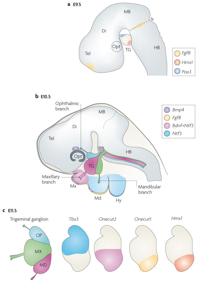
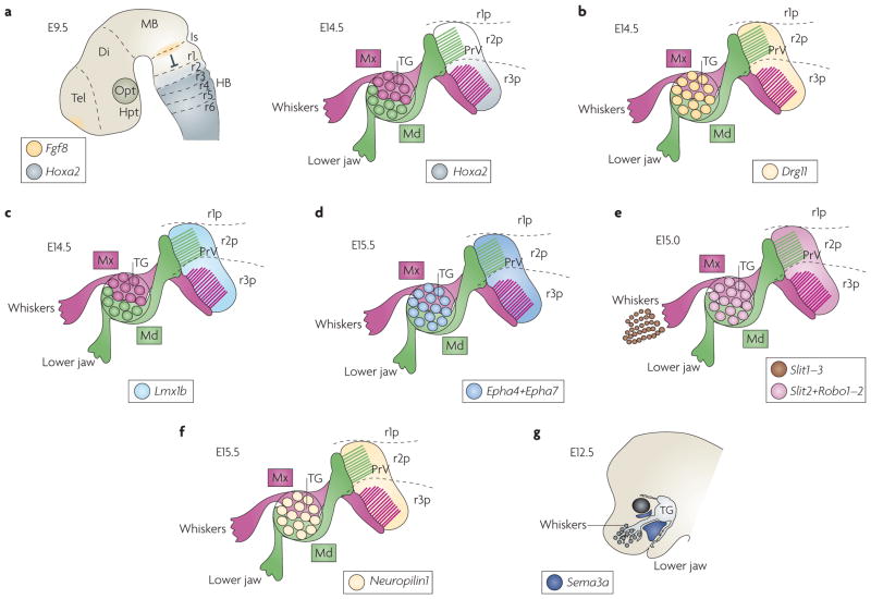
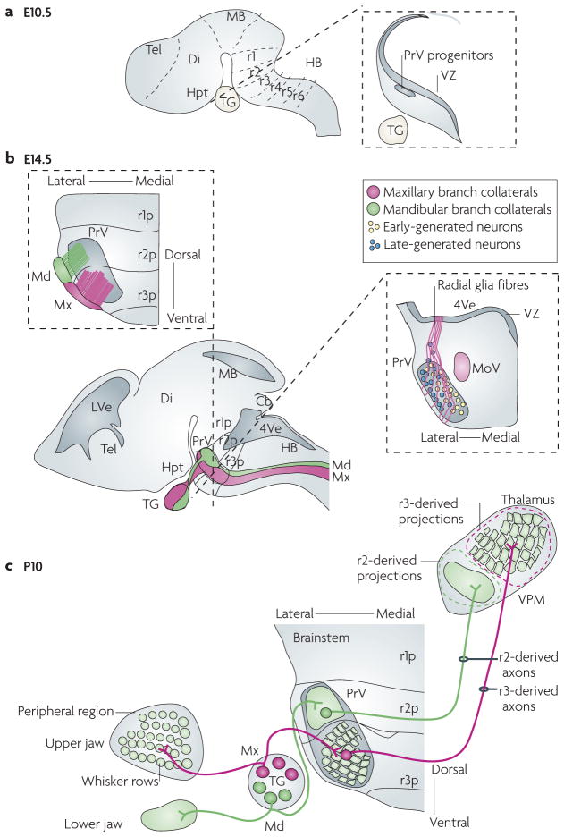
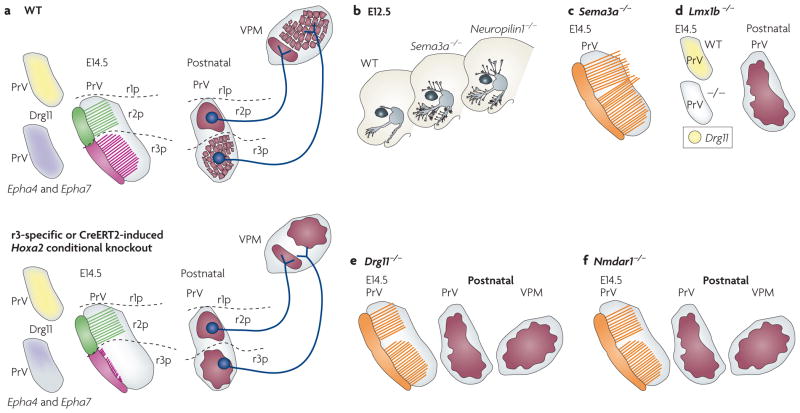
The facial somatosensory map in the cortex is derived from facial representations that are first established at the brainstem level and then serially 'copied' at each stage of the somatosensory pathway. Recent studies have provided insights into the molecular mechanisms involved in the development of somatotopic maps of the face and whiskers in the trigeminal nuclei of the mouse brainstem. This work has revealed that early molecular regionalization and positional patterning of trigeminal ganglion and brainstem target neurons are established by homeodomain transcription factors, the expression of which is induced and maintained by signals from the brain and face. Such position-dependent information is fundamental in transforming the early spatial layout of sensory receptors into a topographic connectivity map that is conferred to higher brain levels.


Conflict of interest statement

Competing interests statement

The authors declare no competing financial interests.

Figures

Figure 1
Figure 1. Trigeminal circuit and face maps in the mouse brain
The ophthalmic (supplying the skin above the eye and forehead), maxillary (supplying the whiskers, upper jaw and lip) and mandibular (supplying the lower jaw and lip) branches of the trigeminal ganglion convey an inverted face map to the brainstem trigeminal nuclei — the rostral principal nucleus (PrV) and the caudal spinal nucleus (SpV). The whiskers and sinus hairs on the snout are innervated by the infraorbital branch of the maxillary nerve (ION). Here, five rows of whiskers (A–E) and the straddle whiskers (α –δ are indicated and colour coded. In the brainstem, radial collaterals emerge from the central trigeminal axons and innervate the PrV and SpV, where they form whisker-specific patterns (barrelettes). In the PrV, the facial map is inverted, with the mandibular fields represented dorsally and maxillary and ophthalmic fields represented ventrally. Similarly, the whisker rows A–E are represented in an inverted fashion. Trigeminothalamic axons from the PrV (lemniscal pathway,) project to the contralateral dorsomedial part of the ventral posteromedial nucleus (VPM) in the thalamus, where the whisker-related neural modules (barreloids) and face map again shift their orientation. SpV neurons project instead to the posteromedial (POm) nucleus (paralemniscal pathway,) and to the ventrolateral VPM (extralemniscal pathway; not shown here for simplicity). Finally, thalamocortical axons from the VPM convey the facial map and whisker patterning to the somatosensory cortex (S1), where barrels form. Figure is modified, with permission, from REF. © (2006) American Association for the Advancement of Science.
Figure 2
Figure 2. Positional molecular patterning of trigeminal ganglion divisions
a | Diagram of the brain at embryonic day (E) 9.5 showing the trigeminal ganglion (TG), its position relative to the hindbrain (HB) and isthmic organizer (Is). Wnt (not shown) and fibroblast growth factor 8 (FGF8) signalling molecules originating at the isthmus cooperate for the spatially restricted induction of the homeodomain transcription factor paired box protein PAX3 in the ophthalmic placode and in progenitors that will become ophthalmic neurons,. Moreover, expression of the Hmx1 homeodomain transcription factor is restricted to the ventral, mandibular trigeminal ganglion division. b | Expression patterns of signalling molecules in the developing trigeminal peripheral targets at E10.5. Neurotrophin 3 (Ntf3) and brain-derived neurotrophic factor (Bdnf) are expressed by both the target epithelium and the mesenchyme through which the TG axons extend. Bone morphogenetic protein 4 (Bmp4) is expressed in regions adjacent to ophthalmic and maxillary regions, although not in mandibular axons, and differentially regulates positionally restricted expression of homeodomain transcription factors in TG neurons. c | Differential homeodomain gene expression in TG divisions at E11.5. T box family transcription factor Tbx3 is restricted to dorsal (ophthalmic and maxillary) TG neurons, whereas Onecut1 (also known as Oc1 and Hnf6) and Hmx1 are expressed by ventral (mandibular) TG neurons, and Onecut2 (also known as Oc2 and Hnf6b) is expressed in the mandibular and ventral half of the maxillary divisions. BMP4 is required to maintain expression of Tbx3 in dorsal TG neurons while suppressing the transcription of Onecut1, Onecut2 and Hmx1 (REF. 44). Di, diencephalon; Hy, hyoid arch; MB, midbrain; Md, mandibular region of the first branchial arch; Mx, maxillary region of the first branchial arch; Opt, optic cup; Tel, telencephalon.
Figure 3
Figure 3. Late expression patterns of homeodomain transcription factors and guidance molecules in the developing TG and PrV
a | In the rostral principal nucleus (PrV) at embryonic day (E) 14.5 (right panel), homeobox A2 (Hoxa2) is differentially expressed in rhombomere 2 (r2)- or r3-derived postmitotic progenies. Hoxa2 is expressed at high levels in the ventral r3-derived portion (r3p), but is barely expressed in the dorsal r2-derived portion (r2p) of the PrV. By contrast, in the hindbrain at E9.5 (left panel), the anterior-most expression domain of Hoxa2 is in r2. Fibroblast growth factor 8 (FGF8)-mediated repression from the isthmus (Is) prevents Hox gene expression in r1 (and perhaps allows only low Hoxa2 expression levels in r2). b | At E14.5, the paired homeodomain factor dorsal root ganglion 11 (Drg11; also known as Drgx) is expressed throughout the PrV and in trigeminal ganglion cells. c | At E14.5, the LIM homeodomain transcription factor Lmx1b is expressed in PrV but not in trigeminal ganglia (TG). d | Ephrin receptor A4 (Epha4) and Epha7 are expressed in the PrV and TG at E15.5 (REF. 72). e | Expression of the Slit ligands and Robo receptors. Slit1, Slit2 and Slit3 are expressed in whisker follicles and Slit2, Robo1 and Robo2 are expressed in both PrV and TG,. f | Expression of the receptor Neuropilin1 at E14.5 occurs in both the PrV and TG. g | The drawing represents X-gal staining at E12.5 of transgenic mice with heterozygous Lacz-knock-in into the semaphorin 3A gene (Sema3a). The trigeminal nerve seldom invades the Sema3a-expressing area. Di, diencephalon; HB, hindbrain; Hpt, hypothalamus; MB, midbrain; Md, mandibular branch of the trigeminal nerve; Mx, maxillary branch of the trigeminal nerve; Opt, optic cup; Tel, telencephalon.
Figure 4
Figure 4. Relationship between rhombomere progenies and PrV somatotopy
a | The developing mouse brain at embryonic day (E) 10.5. The left drawing shows rhombomere compartments of the hindbrain. The right illustration depicts a representative coronal section through the rhombomere 2 (r2) region of the hindbrain. At this stage, rostral principal nucleus (PrV) progenitors first emerge from the ventricular zone (VZ). b | Lateral view of the mouse brain at E14.5 (bottom left panel). Central axons of the trigeminal nerve — the mandibular branch (shown in green) and the maxillary branch (shown in pink) — enter the hindbrain, form the trigeminal tract and innervate the PrV. The upper section shows the somatotopic relationship between the r2- and r3-derived progenies contributing to the PrV and their targeting by distinct trigeminal nerve branches. Specifically, mandibular-branch axons mainly arborize into the r2-derived portion of PrV, but not the r3-derived component. Conversely, r3-derived neurons receive selective collateral input from whisker-related, but not mandibular, maxillary afferents. The right section shows the migration of PrV neurons from the VZ along radial fibres and their ‘inside-out’ distribution in the PrV, with neurons that are formed early settling more medially than neurons that are formed late. c | Topography of the trigeminal circuit at postnatal day 10. The illustration shows the topography of afferent and efferent axonal connections of the PrV nucleus in relation to rhombomere-derived neuronal progenies (r1p–r3p). The rhombomere-specific spatial segregation of neurons in the mature PrV underlies the parcelling of mandibular and maxillary (whisker-related) segments of the face map. Cb, cerebellum; Di, diencephalon; HB, hindbrain; Hpt, hypothalamus; LVe, lateral ventricle; MB, midbrain; MoV, motor trigeminal nucleus; Tel, telencephalon; 4Ve, fourth ventricle; VPM, ventral posterior medial nucleus .
Figure 5
Figure 5. Phenotypes of the trigeminal system in mutant mice
a The trigeminal system at embryonic day (E) 14.5. The upper panel represents the wild type (WT) and the lower panel represents the similar phenotypes of rhombomere 3 (r3)-specific or temporally induced homeobox A2 (Hoxa2) conditional mutants. The left drawings show the expression of dorsal root ganglion 11 (Drg11; also known as Drgx), ephrin receptor A4 (Epha4) and Epha7 in the rostral principal nucleus (PrV). The middle drawings show the collaterals of trigeminal mandibular branches (shown in green) and whisker-related maxillary branches (shown in pink) in the PrV in relation to the spatial restriction of rhombomere progenies. The right drawings show the projection of the r2- and r3- derived axons and the somatotopic map of the PrV and ventral posteromedial nucleus (VPM) at postnatal stages. Inactivation of Hoxa2 in r3 or before collateral formation specifically impairs collateral formation from incoming trigeminal ganglion (TG) whisker-related afferents and arborization onto PrV neurons, resulting in lack of barrelette pattern. Mutant PrV neuron projections can normally be traced to the contralateral VPM, but display topographic mapping defects that correlate with reduced Epha4 and Epha7 expression in prenatal PrV and result in lack of barreloids. b | Schematic representation of the peripheral distribution of the trigeminal branches in semaphorin 3A (Sema3a)- and Neuropilin1-knockout mice. SEMA3A and its receptor neuropilin have a role in restricting peripheral (shown in grey) and central (shown in orange) trigeminal axons to specific routes but are not involved in whisker-related patterning in the PrV. c | Phenotypes of the central branch of the trigeminal nerve in Sema3a-knockout mice. Note that the central trigeminal ganglion axon collaterals (shown in orange) pass beyond the PrV. d | The trigeminal system in a LIM homeobox transcription factor 1β (Lmx1b) mutant. The left drawings show loss of Drg11 expression in the mutant PrV at E14.5. The right drawing shows the loss of somatotopic map in the postnatal mutant PrV. e,f | The trigeminal system in Drg11- and Nmdar1 (N-methyl 3-aspartate receptor 1) knockout mice, respectively. The PrV phenotypes of Lmx1B, Drg11 and Nmdar1 knockouts are similar in that they all lack barrelettes.

Similar articles

Cited by

References

    1. Penfield W, Boldrey E. Somatic motor and sensory representation in the cerebral cortex of man as studied by electrical stimulation. Brain. 1937;60:389–443.
    1. Belford GR, Killackey HP. The development of vibrissae representation in subcortical trigeminal centers of the neonatal rat. J Comp Neurol. 1979;188:63–74. - PubMed
    1. Ma PM. The barrelettes — architectonic vibrissal representations in the brainstem trigeminal complex of the mouse. I Normal structural organization. J Comp Neurol. 1991;309:161–199. - PubMed
    1. Ma PM. Barrelettes — architectonic vibrissal representations in the brainstem trigeminal complex of the mouse. II Normal post-natal development. J Comp Neurol. 1993;327:376–397. - PubMed
    1. Ma PM, Woolsey TA. Cytoarchitectonic correlates of the vibrissae in the medullary trigeminal complex of the mouse. Brain Res. 1984;306:349–374. This report coined the term ‘barrelettes’ for the whisker-specific neural modules. - PubMed

Publication types