. 2010 Feb 22;5(2):e9342.
doi: 10.1371/journal.pone.0009342.

Mechanistic issues of the interaction of the hairpin-forming domain of tBid with mitochondrial cardiolipin

Affiliations

Mechanistic issues of the interaction of the hairpin-forming domain of tBid with mitochondrial cardiolipin

François Gonzalvez et al. PLoS One. .

Abstract

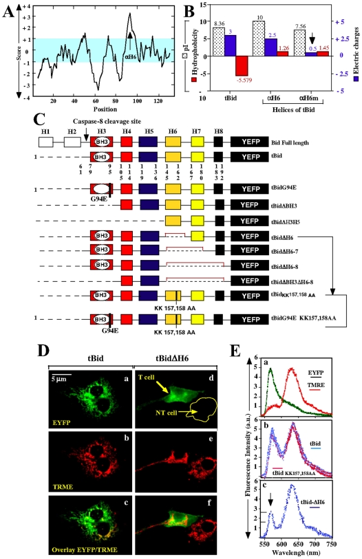
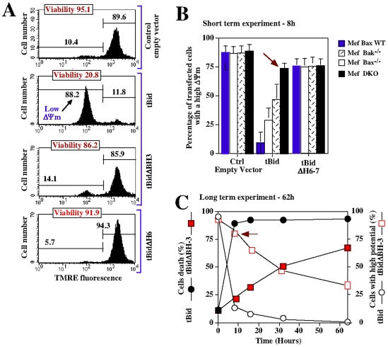
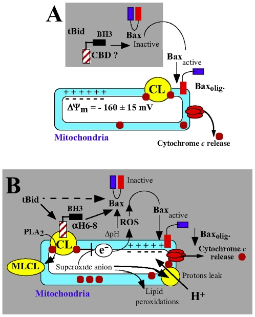
Background: The pro-apoptotic effector Bid induces mitochondrial apoptosis in synergy with Bax and Bak. In response to death receptors activation, Bid is cleaved by caspase-8 into its active form, tBid (truncated Bid), which then translocates to the mitochondria to trigger cytochrome c release and subsequent apoptosis. Accumulating evidence now indicate that the binding of tBid initiates an ordered sequences of events that prime mitochondria from the action of Bax and Bak: (1) tBid interacts with mitochondria via a specific binding to cardiolipin (CL) and immediately disturbs mitochondrial structure and function idependently of its BH3 domain; (2) Then, tBid activates through its BH3 domain Bax and/or Bak and induces their subsequent oligomerization in mitochondrial membranes. To date, the underlying mechanism responsible for targeting tBid to mitochondria and disrupting mitochondrial bioenergetics has yet be elucidated.

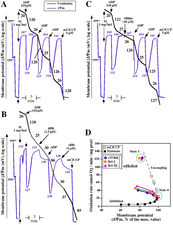
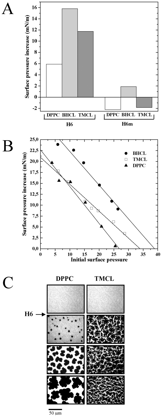
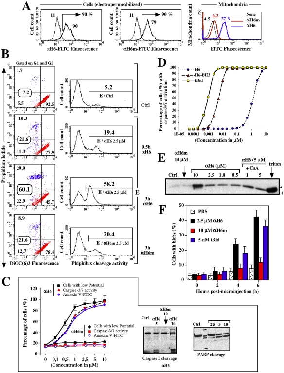
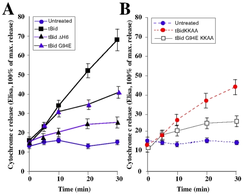
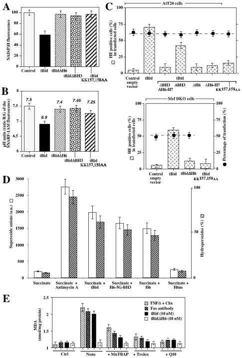
Principal findings: The present study investigates the mechanism by which tBid interacts with mitochondria issued from mouse hepatocytes and perturbs mitochondrial function. We show here that the helix alphaH6 is responsible for targeting tBid to mitochondrial CL and disrupting mitochondrial bioenergetics. In particular, alphaH6 interacts with mitochondria through electrostatic interactions involving the lysines 157 and 158 and induces an inhibition of state-3 respiration and an uncoupling of state-4 respiration. These changes may represent a key event that primes mitochondria for the action of Bax and Bak. In addition, we also demonstrate that tBid required its helix alphaH6 to efficiently induce cytochrome c release and apoptosis.

Conclusions: Our findings provide new insights into the mechanism of action of tBid, and particularly emphasize the importance of the interaction of the helix alphaH6 with CL for both mitochondrial targeting and pro-apoptotic activity of tBid. These support the notion that tBid acts as a bifunctional molecule: first, it binds to mitochondrial CL via its helix alphaH6 and destabilizes mitochondrial structure and function, and then it promotes through its BH3 domain the activation and oligomerization of Bax and/or Bak, leading to cytochrome c release and execution of apoptosis. Our findings also imply an active role of the membrane in modulating the interactions between Bcl-2 proteins that has so far been underestimated.


Conflict of interest statement

Competing Interests: The authors have declared that no competing interests exist.

Figures

Figure 1
Figure 1. The helix αH6 targets tBid to the mitochondria.
(A) and (B) Computer-based analysis of the biophysical properties of Bid. (A) Kite & Doolittle profile of Bid (PROSTCALE, Swiss Institute of Bioinformatics). (B) Comparison of the isoelectric points (pI), hydrophobicities and charges of tBid, αH6 and αH6m. (C) Schematic representation of tBid-EYFP mutants. (D) and (E) CV-1 cells were transfected with plasmids endoding tBid-EYFP mutants in presence of 10 µM of Bok-D to inhibit caspases activation and subsequent cell death. 24 h later, cells were stained with 20 nM of the mitochondrial potential probe TMRE and the localization of the tBid-EYFP mutants was determined using confocal microscopy (D) and microspectrofluorometry analysis (E).
Figure 2
Figure 2. The helix αH6 affects mitochondrial bioenergetics.
Purified mice liver mitochondria (M) were incubated in respiratory buffer (0.33 mg/ml). Oxygen consumption (Voxidation, black line) and mitochondrial potential (ΔΨm, bleu line) were monitored using a Clark-type electrode coupled to a tetraphenylphosphonium (TPP+) cation-sensitive electrode, as described previously . 10 mM of succinate was added as oxidizable substrate (malonate is 1 mM). ADP was added to 110 µM and mCCCP was added to 10 µM. The numbers along the traces gives the values of oxidation rates in nmol O2/min/mg protein (Black) and mitochondrial potential in mV (Blue) (A) Oxygen consumption and potential of mitochondria oxidizing succinate. (B) and (C) Effect of the addition αH6 and αH6m on the oxygen consumption and mitochondrial potential. Each panels (A), (B) and (C) is representative of 3 independent experiments. (D) Representation of the effect of αH6 on oxidation rate and potential of mitochondria isolated from control, Bcl-2 and Bcl-XL transgenic mice, as compared to uncoupling and inhibition of the mitochondrial respiration. Dotted black line shows the effect of the uncoupler CCCP, and the solid black line represents the inhibition of succinate-respiration by the specific inhibitor of complex II, malonate.
Figure 3
Figure 3. The helix αH6 specifically inserts into CL-monolayers through electrostatic interactions and reorganizes them into microdomains.
Dipalmitoylphosphatidylcholine (DPPC), bovine heart CL (BHCL) and tetramyristoyl CL (TMCL) were spread at an initial surface pressure of 20 mN/m. αH6 and αH6m (1 µM) were injected into the subphase of these lipids monolayers. (A) The surface pressure changes (B) and epifluorescence images (C) were recorded as described previously (Gonzalvez et al 2005, CDD).
Figure 4
Figure 4. Electroporation and microinjection of αH6 induces apoptosis in Jurkat cells.
Jurkat cells were electroporated with 2.5 µM of αH6-FITC and αH6m-FITC peptides, as described previously (Gabriel 2003). The efficiency of peptides electroporation into these cells was determined by measuring the FITC fluorescence using flow cytometry (A). Mitochondria were isolated from these cells, and analyze by flow cytometry described previously . (B and C) Effect of αH6 and αH6m electroporation (2.5 µM) on mitochondrial potential, caspases 3/7 activation and cell viability in Jurkat cells. (B) Left panels: dot plot of DIOC(6)3 vs PI staining cells. Right panels: caspases 3/7 activities by cleavage of the Phiphilux substrate. E refers to electroporated cells. (C) Left pannels: dose response of αH6 and αH6m on mitochondrial potential, caspases 3/7 activation and apoptosis. Right panels: Caspase-3 and PARP cleavage were analyzed by Western Blotting. (D) Dose response curves of caspases 3/7 activation induced by electropermeabilization of tBid, αH6-BH3, and αH6 in Jurkat cells. (E) Jurkat cells were microinjected with tBid, αH6 and αH6m and the percentage of cell blebbing was measured by microscopy (F). Data are representative of 10 independant measurements (n = 10).
Figure 5
Figure 5. αH6 and BH3 domains are both required for tBid-induced apoptosis.
(A) Jurkat cells were transfected with either empty vector control or with plasmids encoding tBid, tBidΔBH3 and tBidΔH6. 12 h latter mitochondrial membrane potential ΔΨm (TMRE) and cell viability (PI) were assessed by FACS analysis. Each panels is representative of 3 independent experiments. (B) Wild-type, Bax+/+, Bax−/−, Bak−/− or double-knockout (DKO) Mefs cells were transfected with either empty vector control or with plasmids encoding tBid and tBidΔH6. After 8 h, green cells were analyzed by FACS for mitochondrial potential using TMRE. The red arrow indicates the ΔΨm in DKO Mefs. (C) Mefs cells were transfected with plasmids encoding tBid and tBidΔBH3, and the kinetics of mitochondrial depolarization and cell death were measured over 60 h by FACS analysis.
Figure 6
Figure 6. αH6 and BH3 domains are both required for tBid cytochrome c release activity.
(A) and (B) Jurkat cells were electroporated with plasmids encoding tBid, tBidΔH6, tBidG94E, tBidKKAA and tBidG94EKKAA and the kinetics of cytochrome c release in the cytosolic fractions were detected by ELISA.
Figure 7
Figure 7. tBid required its helix αH6, but not its BH3 domain, to induce superoxide anion production and mitochondrial lipid peroxidation.
(A) and (B) Wild-type hepatocytes were transfected with a control vector (control) or plasmids encoding tBid, tBidΔH6, tBidΔBH3 and tBidKKAA and NAD(P)H and SNARF-1AM fluorescence (pH indicator) were measured by FACS. Data are given as % of the control ± SD. pH units were determined using a calibration curve generated using nigericin-permeabilized cells kept in buffer of different pH values. (C) AtT20 cells or DKO Mefs were transfected with empty vector (control) or plasmids encoding tBid, tBidΔH6, tBidΔBH3, tBid tBidKKAA, tBidΔH6–H7, tBidΔBH3ΔH6H7. Cells were then stained with hydroethidine (HE, Invitrogen/Molecular probes) to measure superoxide anion production. The percentages of transfected cells are indicated by the dotted line. (D) Purified mice liver mitochondria were energized using succinate (+ rotenone) and treated using 10 nM tBid, H6-5G-BH4, αH6 and αH6m. Mitochondrial superoxide anion production was measured using HE (a.u.  =  arbitrary units) whereas hydroperoxide was measured using amplex red. (E) Wild-type hepatocytes were treated with TNFα/cycloheximide, anti-Fas antibody or transfected with a plasmids encoding tBid and tBidΔH6. Mitochondrial lipid peroxidation was measured by FACS using MDA. We used the antioxidants trolox (2 mM), MnTBAP (1 mM) and MitoQ10 (1 µM).
Figure 8
Figure 8. tBid, a bifunctional molecule.
(A) Current model of the BH3-dependant function of tBid. tBid interacts through its BH3 domain and directly activates Bax, which undergoes conformational changes that induce the exposure of its N-terminal domains. This results in the stable insertion and subsequent oligomerization of Bax in the mitochondrial outer membrane leading to the release of cytochrome c and apoptosis. This model highlights the importance of protein-protein interactions between tBid and Bax. (B) Refined model of the pro-apoptotic function of tBid: importance of tBid/CL interactions. First, tBids binds to CL present at the contact sites via its helix αH6 and destabilizes the mitochondrial membrane. This may affect the activity of the electron transport chain complexes and lead to an acidification of the cytosol, mitochondrial ROS production and mitochondrial lipid peroxidation. This environment may prime the activation of Bax and/or Bak. Then, tBid interacts through its BH3 domain with Bax and/or Bak to promote their oligomerization and subsequently induce cytochrome c release and apoptosis.

Similar articles

Cited by

References

    1. Cory S, Adams JM. The Bcl2 family: regulators of the cellular life-or-death switch. Nat Rev Cancer. 2002;2:647–656. - PubMed
    1. Petros AM, Olejniczak ET, Fesik SW. Structural biology of the Bcl-2 family of proteins. Biochim Biophys Acta. 2004;1644:83–94. - PubMed
    1. Muchmore SW, Sattler M, Liang H, Meadows RP, Harlan JE, et al. X-ray and NMR structure of human Bcl-xL, and inhibitor of programmed cell death. Nature. 1996;381:335–341. - PubMed
    1. Adams JM, Cory S. The Bcl-2 protein family: arbiters of cell survival. Science. 1998;281:1322–1326. - PubMed
    1. Letai A, Bassik M, Walensky L, Sorcinelli M, Weiler S, et al. Distinct BH3 domains either sensitize or activate mitochondrial apoptosis, serving as prototype cancer therapeutics. Cancer Cell. 2002;2:183. - PubMed

Publication types

MeSH terms