

Metastatic lung adenocarcinoma in a 20-year-old patient
- PMID: 20179804
- PMCID: PMC2826778
- DOI: 10.3747/co.v17i1.543
Metastatic lung adenocarcinoma in a 20-year-old patient
Abstract
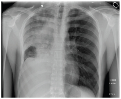
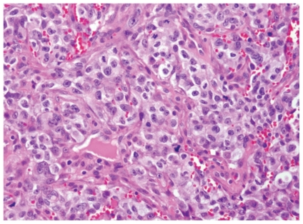
Lung cancer is rare disease in patients under 25 years of age. It typically occurs in older patients with a history of tobacco use. This case concerns a 20-year-old man with no history of tobacco use who complained of several months of cough and lower back pain and an 11.3-kg weight loss. He was treated for pneumonia after a chest radiograph showed total opacification of the right lung. Computed tomography imaging subsequently revealed a superior right hilar mass and mediastinal lymphadenopathy. Further imaging studies showed diffuse metastatic disease. Mediastinal biopsy showed poorly differentiated epithelioid tumour with desmoplastic stromal reaction, neutrophil infiltration, and squamous differentiation. Tissue immunostaining confirmed a non-small-cell lung cancer. Unfortunately, despite aggressive therapy, the patient's disease progressed, and he died within 9 months. In this paper, we hope to illustrate the unique challenges in diagnosing and treating young patients with metastatic lung cancer.
Keywords: Metastatic lung cancer; non-small-cell lung cancer; young patients.
Figures

Similar articles
-
[Pulmonary mucosa-associated lymphoid tissue lymphoma concurrent with lung squamous cell carcinoma: a case report and literature review].Zhonghua Jie He He Hu Xi Za Zhi. 2020 Dec 12;43(12):1071-1076. doi: 10.3760/cma.j.cn112147-20200729-00859. Zhonghua Jie He He Hu Xi Za Zhi. 2020. PMID: 33333642 Review. Chinese.
-
[Mediastinal and hilar lymph node of cancer unknown origin: 3 case reports].Nihon Kokyuki Gakkai Zasshi. 1999 Jan;37(1):72-7. Nihon Kokyuki Gakkai Zasshi. 1999. PMID: 10087881 Review. Japanese.
-
Sister Mary Joseph's nodule as a presenting sign of internal malignancy.Skinmed. 2006 Sep-Oct;5(5):256-8. doi: 10.1111/j.1540-9740.2006.04826.x. Skinmed. 2006. PMID: 16957443
-
Rare coexistence of sarcoidosis and lung adenocarcinoma.Respir Med Case Rep. 2014 Mar 15;12:4-6. doi: 10.1016/j.rmcr.2013.12.008. eCollection 2014. Respir Med Case Rep. 2014. PMID: 26029525 Free PMC article.
-
A Rare Case of Metastatic Desmoplastic Small Round Cell Tumour: Diagnosis and Management.Case Rep Oncol Med. 2015;2015:925453. doi: 10.1155/2015/925453. Epub 2015 Aug 9. Case Rep Oncol Med. 2015. PMID: 26347069 Free PMC article.
Cited by
-
Lung adenocarcinoma presumed to be Pott's disease in a 28-year-old patient: A case report and review of literature.Surg Neurol Int. 2019 Oct 18;10:208. doi: 10.25259/SNI_403_2019. eCollection 2019. Surg Neurol Int. 2019. PMID: 31768288 Free PMC article.
References
-
- Jemal A, Siegal R, Ward E, Hao Y, Xu J, Thun MJ. Cancer statistics, 2009. CA Cancer J Clin. 2009;59:225–49. - PubMed
-
- Sun S, Schiller JH, Gazdar AF. Lung cancer in never smokers— a different disease. Nat Rev Cancer. 2007;7:778–90. - PubMed
-
- United States, National Institutes of Health, National Cancer Institute, Surveillance Epidemiology and End Results (seer) Cancer Statistics Review, 1975–2006. Bethesda, MD: National Cancer Institute, Cancer Statistics Branch; 2009. [Available online at: seer.cancer.gov/csr/1975_2006/browse_csr.php?section=15&page=sect_15_tab...; cited July 13, 2009]
-
- Dishop MK, Kuruvilla S. Primary and metastatic lung tumors in the pediatric population: a review and 25-year experience at a large children’s hospital. Arch Pathol Lab Med. 2008;132:1079–103. - PubMed
LinkOut - more resources
Full Text Sources
