Review
. 2010 Mar;39(3):1036-48.
doi: 10.1039/b909900j. Epub 2010 Feb 1.

Fundamentals of microfluidic cell culture in controlled microenvironments

Affiliations
Review

Fundamentals of microfluidic cell culture in controlled microenvironments

Edmond W K Young et al. Chem Soc Rev. 2010 Mar.

Abstract

Microfluidics has the potential to revolutionize the way we approach cell biology research. The dimensions of microfluidic channels are well suited to the physical scale of biological cells, and the many advantages of microfluidics make it an attractive platform for new techniques in biology. One of the key benefits of microfluidics for basic biology is the ability to control parameters of the cell microenvironment at relevant length and time scales. Considerable progress has been made in the design and use of novel microfluidic devices for culturing cells and for subsequent treatment and analysis. With the recent pace of scientific discovery, it is becoming increasingly important to evaluate existing tools and techniques, and to synthesize fundamental concepts that would further improve the efficiency of biological research at the microscale. This tutorial review integrates fundamental principles from cell biology and local microenvironments with cell culture techniques and concepts in microfluidics. Culturing cells in microscale environments requires knowledge of multiple disciplines including physics, biochemistry, and engineering. We discuss basic concepts related to the physical and biochemical microenvironments of the cell, physicochemical properties of that microenvironment, cell culture techniques, and practical knowledge of microfluidic device design and operation. We also discuss the most recent advances in microfluidic cell culture and their implications on the future of the field. The goal is to guide new and interested researchers to the important areas and challenges facing the scientific community as we strive toward full integration of microfluidics with biology.


Figures

Fig. 1
Fig. 1
The cell microenvironment consists of physical, biochemical, and physicochemical factors. For example, the endothelium that lines blood vessels is exposed to hemodynamic shear stress (external physical force) that stimulates a biochemical response, releasing nitric oxide (NO). NO diffuses to neighboring smooth muscle cells (SMCs), where it regulates cell contraction and relaxation. The gradient of diffused NO affects nearby SMCs more than distant SMCs. Endothelial cells are anchored to the basement membrane, while SMCs are anchored to the extracellular matrix of the interstitium, both via integrins that act as sensors and transducers of physical force. Local physicochemical properties ensure proper regulation of both physical and biochemical mechanisms.
Fig. 2
Fig. 2
A fully automated PDMS-based microfluidic cell culture system consisting of 96 individually addressable cell culture chambers (blue dye), on-chip peristaltic pumping (lower right inset), and multiplexing capabilities that allow different mixtures of reagents to be formulated. Reprinted with permission from Gomez-Sjoberg et al Copyright 2007 American Chemical Society.
Fig. 3
Fig. 3
Passive pumping relies on surface tension of small droplets to pump fluid from inlet to outlet. A smaller drop of radius Ri has an internal pressure Pi greater than the pressure in a larger drop of radius Ro because of Laplace’s law (ΔP = 2γ/R), where ΔP is the pressure difference across the liquid–air interface of the droplet, and γ is the surface tension at the interface.
Fig. 4
Fig. 4
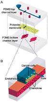
PDMS-based microfluidic device containing a porous polyester membrane for supporting growth of endothelial cells. Device was used to study adhesion of cancer cells on the endothelium in the presence of chemokines released on the basolateral side of the endothelium. (Open access: Song et al., PLoS One, 2009, 4(6); 5756.)
Fig. 5
Fig. 5
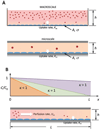
Effective culture time (ECT) and critical perfusion rate (CPR). (A) Macroscale static cultures have larger h and therefore a larger Damkohler number compared to static microscale cultures. Because substrate uptake time scales dominate at the microscale, media must be replenished sooner based on the change in media height. (B) To ensure all cells in a microfluidic culture are being replenished sufficiently with perfused media, perfusion rate Um must be large enough to displace exhausted media (κ > 1). CPR is defined as the perfusion rate where κ = 1. This can be determined by dividing channel length with ECT.
Fig. 6
Fig. 6
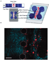
(A) Microfluidic coculture device for three-dimensional (3D) microenvironments. (B) The microfluidic coculture device allows imaging of collagen fibers (blue) via Second Harmonic Generation. Matrix remodeling is observed as cells cluster at the boundary of the gels. Scale bar = 100 µm. (Huang et al.—reproduced by permission of The Royal Society of Chemistry.)
Fig. 7
Fig. 7
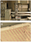
Interfacing microchannel arrays with automated liquid handling systems for high-throughput biological studies. (A) Four-channel automated liquid handling system (Gilson Quad-Z 215 Liquid Handler, Middleton, WI, USA) interfaced with microchannel plate (white box). (B) Array of 192 microchannels in standard microtiter plate format. Straight microchannels contain access ports to allow passive pumping.

Similar articles

Cited by

References

    1. Beebe DJ, Mensing GA, Walker GM. Annu. Rev. Biomed. Eng. 2002;4:261–286. - PubMed
    1. Paguirigan AL, Beebe DJ. BioEssays. 2008;30:811–821. - PMC - PubMed
    1. Walker GM, Zeringue HC, Beebe DJ. Lab Chip. 2004;4:91–97. - PubMed
    1. Andersson H, van den Berg A. Sens. Actuators, B. 2003;92:315–325.
    1. El-Ali J, Sorger PK, Jensen KF. Nature. 2006;442:403–411. - PubMed

Publication types