

Removal of an oral squamous cell carcinoma including parts of osseointegrated implants in the marginal mandibulectomy. A case report
- PMID: 20180142
- PMCID: PMC2988214
- DOI: 10.1007/s10006-010-0208-y
Removal of an oral squamous cell carcinoma including parts of osseointegrated implants in the marginal mandibulectomy. A case report
Abstract
Purpose: The incidence of oral squamous cell carcinomas (OSCC) arising around dental implants will increase because of the rising popularity of dental implants. In this case, a novel surgical treatment of an OSCC in the vicinity of endosseous implants is reported.
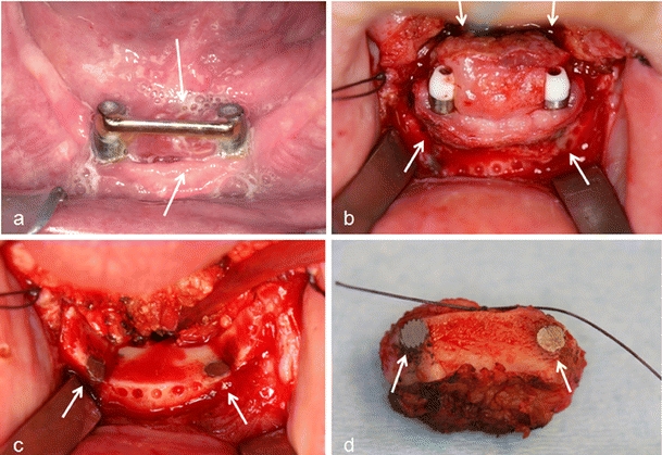
Materials and methods: In a 69-year-old woman, a recurrent OSCC (cT₂N₀M₀) developed in the floor of the mouth extending to the attached keratinized peri-implant mucosa of both interforaminal-placed dental implants. Radiographically, no bone invasion could be observed.
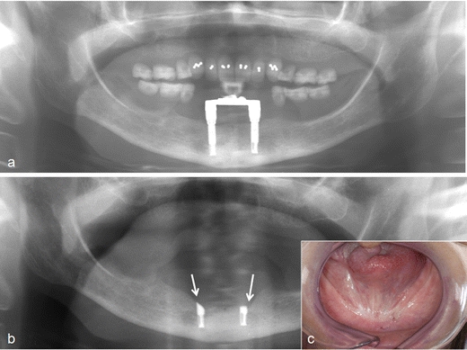
Results: To radically remove the tumor, a marginal mandibulectomy was performed including the cranial parts of both dental implants by cutting them into two parts. Three years after tumor resection and one year after reimplantation, the patient is disease free and has a good oral function.
Conclusions: In case of an OSCC, traditional bone and soft margins for oncologic safety are 1.0 cm. If a dental implant is present within this safety zone, on condition, there is no massive bone invasion, and the original mandible has sufficient vertical height; a marginal mandibulectomy including part of the implants can be considered.
Figures

Similar articles
-
[On the indications for and morbidity of segmental resection of the mandible for squamous cell carcinoma in the lower oral cavity].Mund Kiefer Gesichtschir. 2005 May;9(3):137-42. doi: 10.1007/s10006-005-0607-7. Mund Kiefer Gesichtschir. 2005. PMID: 15834743 German.
-
The effect of different transplanted soft tissues on bone resorption around loaded endosseous implants in patients after oral tumor surgery.Int J Oral Maxillofac Implants. 1998 Jul-Aug;13(4):554-60. Int J Oral Maxillofac Implants. 1998. PMID: 9714963
-
Marginal and segmental mandibulectomy in patients with oral cancer: a statistical analysis of 106 cases.J Oral Maxillofac Surg. 2003 Nov;61(11):1289-96. doi: 10.1016/s0278-2391(03)00730-4. J Oral Maxillofac Surg. 2003. PMID: 14613085
-
Anterior floor of mouth resection with marginal mandibulectomy.Atlas Oral Maxillofac Surg Clin North Am. 1997 Sep;5(2):37-54. Atlas Oral Maxillofac Surg Clin North Am. 1997. PMID: 11905315 Review. No abstract available.
-
Histopathological Features of Secondary Squamous Cell Carcinoma Around a Dental Implant in the Mandible After Chemoradiotherapy: A Case Report With a Clinicopathological Review.J Oral Maxillofac Surg. 2016 May;74(5):982-90. doi: 10.1016/j.joms.2015.11.004. Epub 2015 Nov 10. J Oral Maxillofac Surg. 2016. PMID: 26679554 Review.
Cited by
-
Is there an association between dental implants and squamous cell carcinoma?Br Dent J. 2016 Nov 18;221(10):645-649. doi: 10.1038/sj.bdj.2016.863. Br Dent J. 2016. PMID: 27857102 Review.
-
Primary radical ablative surgery and fibula free-flap reconstruction for T4 oral cavity squamous cell carcinoma with mandibular invasion: oncologic and functional results and their predictive factors.Eur Arch Otorhinolaryngol. 2017 Jan;274(1):441-449. doi: 10.1007/s00405-016-4219-7. Epub 2016 Jul 20. Eur Arch Otorhinolaryngol. 2017. PMID: 27438536
-
Prosthodontic rehabilitation of patient with marginal mandibular resection using attachment supported prostheses: A clinical report.Contemp Clin Dent. 2014 Jan;5(1):123-6. doi: 10.4103/0976-237X.128690. Contemp Clin Dent. 2014. PMID: 24808712 Free PMC article.
-
Oral squamous cell carcinoma in the vicinity of dental implants.Clin Oral Investig. 2014 Jan;18(1):277-84. doi: 10.1007/s00784-013-0968-5. Epub 2013 Mar 16. Clin Oral Investig. 2014. PMID: 23504205
-
An Update on Implant-Associated Malignancies and Their Biocompatibility.Int J Mol Sci. 2024 Apr 24;25(9):4653. doi: 10.3390/ijms25094653. Int J Mol Sci. 2024. PMID: 38731871 Free PMC article. Review.
References
-
- Schoen PJ, Raghoebar GM, Bouma J, Reintsema H, Burlage FR, Roodenburg JL, Vissink A. Prosthodontic rehabilitation of oral function in head–neck cancer patients with dental implants placed simultaneously during ablative tumor surgery: an assessment of treatment outcomes and quality of life. Int J Oral Maxillofac Surg. 2008;37:8–16. doi: 10.1016/j.ijom.2007.07.015. - DOI - PubMed
-
- Clapp C, Wheeler JC, Martof AB, Levine PA. Oral squamous cell carcinoma in association with dental osseointegrated implants. An unusual occurrence. Arch Otolaryngol Head Neck Surg. 1996;122:1402–1403. - PubMed
Publication types
MeSH terms
Substances
LinkOut - more resources
Full Text Sources
Medical
