

Pathological differential diagnosis of solid-pseudopapillary neoplasm and endocrine tumors of the pancreas
- PMID: 20180245
- PMCID: PMC2828590
- DOI: 10.3748/wjg.v16.i8.1025
Pathological differential diagnosis of solid-pseudopapillary neoplasm and endocrine tumors of the pancreas
Abstract
Aim: To investigate differential points of solid-pseudopapillary neoplasm (SPN) of the pancreas and pancreatic endocrine tumor (PET).
Methods: Ten cases of SPN and fourteen cases of PET were studied in this retrospective study. Clinical and pathologic features, immunostaining reactions and beta-catenin gene mutations were analyzed.
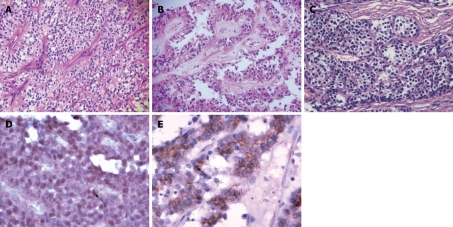
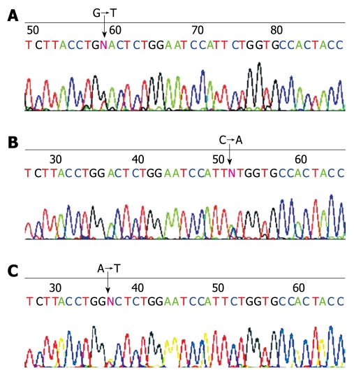
Results: The mean age of SPN patients was 25.6 years and these patients had no specific symptoms. The mean diameter of the tumors was 11.0 cm, 9/10 cases were cystic or a mixture of solid and cystic structures, and there was hemorrhage and necrosis on the cut surface in 8/10 (80%) cases. Characteristic pseudopapillary structure and discohesive appearance of the neoplastic cells were observed in all 10 (100%) cases. The results of immunostaining showed that nuclear expression of beta-catenin and loss of E-cadherin in all the cases, was only seen in SPN. Molecular studies discovered that 9/10 (90%) cases harbored a point mutation of exon 3 in beta-catenin gene. On the other hand, the mean age of PET patients was 43.1 years. Eight of 14 cases presented with symptoms caused by hypoglycemia, and the other 6 cases presented with symptoms similar to those of SPN. The mean size of the tumors was 2.9 cm, most of the tumors were solid, only 3/14 (21%) were a mixture of solid and cystic structures, and macroscopic hemorrhage and necrosis were much less common (3/14, 21%). Histologically, tumor cells were arranged in trabecular, acinar or solid patterns and demonstrated no pseudopapillary structure and discohesive appearance in all 14 (100%) cases. The results of immunostaining and mutation detection were completely different with SPN that membrane and cytoplastic expression of beta-catenin without loss of E-cadherin, as well as no mutation in beta-catenin gene in all the cases.
Conclusion: Both macroscopic and microscopic features of SPN are quite characteristic. It is not difficult to distinguish it from PET. If necessary, immunostaining of beta-catenin and E-cadherin is quite helpful to make the differential diagnosis.
Figures
References
-
- Wick MR, Graeme-Cook FM. Pancreatic neuroendocrine neoplasms: a current summary of diagnostic, prognostic, and differential diagnostic information. Am J Clin Pathol. 2001;115 Suppl:S28–S45. - PubMed
-
- Comper F, Antonello D, Beghelli S, Gobbo S, Montagna L, Pederzoli P, Chilosi M, Scarpa A. Expression pattern of claudins 5 and 7 distinguishes solid-pseudopapillary from pancreatoblastoma, acinar cell and endocrine tumors of the pancreas. Am J Surg Pathol. 2009;33:768–774. - PubMed
-
- Serra S, Chetty R. Revision 2: an immunohistochemical approach and evaluation of solid pseudopapillary tumour of the pancreas. J Clin Pathol. 2008;61:1153–1159. - PubMed
-
- Notohara K, Hamazaki S, Tsukayama C, Nakamoto S, Kawabata K, Mizobuchi K, Sakamoto K, Okada S. Solid-pseudopapillary tumor of the pancreas: immunohistochemical localization of neuroendocrine markers and CD10. Am J Surg Pathol. 2000;24:1361–1371. - PubMed
-
- Kosmahl M, Seada LS, Jänig U, Harms D, Klöppel G. Solid-pseudopapillary tumor of the pancreas: its origin revisited. Virchows Arch. 2000;436:473–480. - PubMed
Publication types
MeSH terms
Substances
LinkOut - more resources
Full Text Sources
Medical
