

Imaging of Abdominal Aortic Aneurysm: the present and the future
- PMID: 20180767
- PMCID: PMC2891873
- DOI: 10.2174/157016110793563898
Imaging of Abdominal Aortic Aneurysm: the present and the future
Abstract
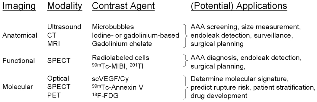
Abdominal Aortic Aneurysm (AAA) is a common, progressive, and potentially lethal vascular disease. A major obstacle in AAA research, as well as patient care, is the lack of technology that enables non-invasive acquisition of molecular/cellular information in the developing AAA. In this review we will briefly summarize the current techniques (e.g. ultrasound, computed tomography, and magnetic resonance imaging) for anatomical imaging of AAA. We also discuss the various functional imaging techniques that have been explored for AAA imaging. In many cases, these anatomical and functional imaging techniques are not sufficient for providing surgeons/clinicians enough information about each individual AAA (e.g. rupture risk) to optimize patient management. Recently, molecular imaging techniques (e.g. optical and radionuclide-based) have been employed to visualize the molecular alterations associated with AAA, which are discussed in this review. Lastly, we try to provide a glance into the future and point out the challenges for AAA imaging. We believe that the future of AAA imaging lies in the combination of anatomical and molecular imaging techniques, which are largely complementary rather than competitive. Ultimately, with the right molecular imaging probe, clinicians will be able to monitor AAA growth and evaluate the risk of rupture accurately, so that the life-saving surgery can be provided to the right patients at the right time. Equally important, the right imaging probe will also allow scientists/clinicians to acquire critical data during AAA development and to more accurately evaluate the efficacy of potential treatments.
Figures
Similar articles
-
Current state of experimental imaging modalities for risk assessment of abdominal aortic aneurysm.J Vasc Surg. 2013 Mar;57(3):851-9. doi: 10.1016/j.jvs.2012.10.097. Epub 2013 Jan 26. J Vasc Surg. 2013. PMID: 23357517 Review.
-
Chronic contained rupture of abdominal aortic aneurysms.J Cardiovasc Surg (Torino). 1996 Feb;37(1):25-8. J Cardiovasc Surg (Torino). 1996. PMID: 8606204
-
Assessing the potential risk of rupture of abdominal aortic aneurysms.Clin Radiol. 2015 Jan;70(1):11-20. doi: 10.1016/j.crad.2014.09.016. Epub 2014 Oct 22. Clin Radiol. 2015. PMID: 25544065 Review.
-
Noninvasive imaging of vascular permeability to predict the risk of rupture in abdominal aortic aneurysms using an albumin-binding probe.Sci Rep. 2020 Feb 24;10(1):3231. doi: 10.1038/s41598-020-59842-2. Sci Rep. 2020. PMID: 32094414 Free PMC article.
-
Surrogate Markers of Abdominal Aortic Aneurysm Progression.Arterioscler Thromb Vasc Biol. 2016 Feb;36(2):236-44. doi: 10.1161/ATVBAHA.115.306538. Epub 2015 Dec 29. Arterioscler Thromb Vasc Biol. 2016. PMID: 26715680 Review.
Cited by
-
Role of molecular imaging with positron emission tomographic in aortic aneurysms.J Thorac Dis. 2017 Apr;9(Suppl 4):S333-S342. doi: 10.21037/jtd.2017.04.18. J Thorac Dis. 2017. PMID: 28540077 Free PMC article. Review.
-
Targeting of microbubbles: contrast agents for ultrasound molecular imaging.J Drug Target. 2018 Jun-Jul;26(5-6):420-434. doi: 10.1080/1061186X.2017.1419362. Epub 2018 Jan 9. J Drug Target. 2018. PMID: 29258335 Free PMC article. Review.
-
Local aortic aneurysm wall expansion measured with automated image analysis.JVS Vasc Sci. 2021 Dec 8;3:48-63. doi: 10.1016/j.jvssci.2021.11.004. eCollection 2022. JVS Vasc Sci. 2021. PMID: 35146458 Free PMC article.
-
Status of diagnosis and therapy of abdominal aortic aneurysms.Front Cardiovasc Med. 2023 Jul 28;10:1199804. doi: 10.3389/fcvm.2023.1199804. eCollection 2023. Front Cardiovasc Med. 2023. PMID: 37576107 Free PMC article. Review.
-
Evaluation of Functionalized Polysaccharide Microparticles Dosimetry for SPECT Imaging Based on Biodistribution Data of Rats.Mol Imaging Biol. 2015 Aug;17(4):504-11. doi: 10.1007/s11307-014-0812-6. Mol Imaging Biol. 2015. PMID: 25537093
References
-
- Belsley SJ, Tilson MD. Two decades of research on etiology and genetic factors in the abdominal aortic aneurysm (AAA)--with a glimpse into the 21st century. Acta Chir Belg. 2003;103:187–196. - PubMed
-
- Jemal A, Siegel R, Ward E, et al. Cancer statistics, 2008. CA Cancer J Clin. 2008;58:71–96. - PubMed
-
- Parodi JC, Palmaz JC, Barone HD. Transfemoral intraluminal graft implantation for abdominal aortic aneurysms. Ann Vasc Surg. 1991;5:491–499. - PubMed
-
- Prinssen M, Verhoeven EL, Buth J, et al. A randomized trial comparing conventional and endovascular repair of abdominal aortic aneurysms. N Engl J Med. 2004;351:1607–1618. - PubMed
-
- Daugherty A, Cassis LA. Mouse models of abdominal aortic aneurysms. Arterioscler Thromb Vasc Biol. 2004;24:429–434. - PubMed
Publication types
MeSH terms
Grants and funding
LinkOut - more resources
Full Text Sources
Medical
