. 2010 May;101(5):1255-60.
doi: 10.1111/j.1349-7006.2010.01497.x. Epub 2010 Jan 18.

Frequent hypermethylation and loss of heterozygosity of the testis derived transcript gene in ovarian cancer

Affiliations

Frequent hypermethylation and loss of heterozygosity of the testis derived transcript gene in ovarian cancer

Haifeng Qiu et al. Cancer Sci. 2010 May.

Abstract

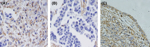
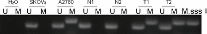
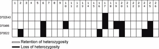
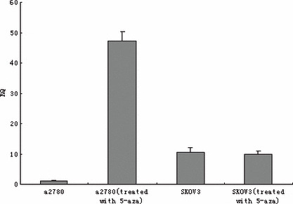
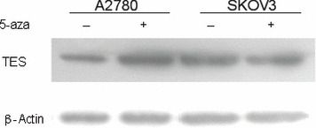
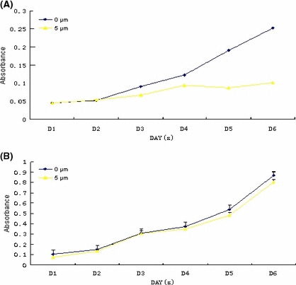
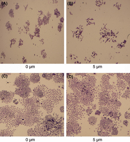
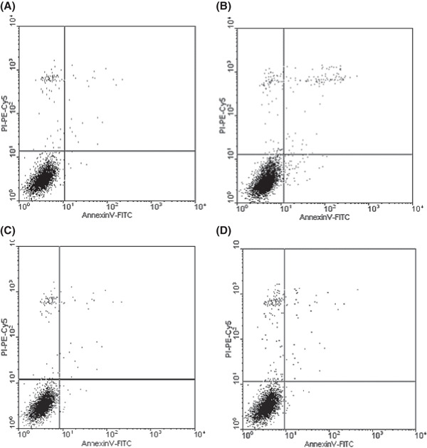
Testis derived transcript (TES) is a candidate tumor suppressor gene located at the human chromosome 7q31, and its function in ovarian cancer is still unknown. Using ovarian cancer cell lines and tissue samples, we demonstrated that both loss of heterozygosity and hypermethylation of the TES gene occurred in ovarian cancer at high frequencies, and there were significant correlations between TES expression and hypermethylation or loss of heterozygosity. We also detected methylation in ovarian cancer cell line A2780 after treatment with 5-aza-2-deoxycytidine. The expression level of TES was enormously up-regulated, then caused changes to the biological behaviors of A2780 cells: cell growth properties were greatly impaired, colony formatting abilities were suppressed to very low levels, and the apoptosis rate was highly raised compared to the control group. Our findings suggest that the TES gene functions as a tumor suppressor gene and is frequently silenced by hypermethylation and loss of heterozygosity in ovarian cancers.


Figures

Figure 1
Figure 1
Testis derived transcript (TES) expression in normal ovary and ovarian cancer tissues. (A) Positive staining of TES protein in ovarian cancer. (B) TES expression was lost in ovarian cancer. (C) Normal ovarian tissue was stained positively.
Figure 2
Figure 2
Methylation analysis of testis derived transcript in ovarian cancers. C, cancer samples; M, methylated; M. Sss I, positive control; N, normal ovary tissues; U, unmethylated. (DNA modificated by CpG methyltransferase M. Sss I.)
Figure 3
Figure 3
Loss of heterozygosity in ovarian cancer.
Figure 4
Figure 4
RT‐PCR analysis of testis derived transcript with or without 5‐aza‐2‐deoxycytidine treatment in A2780 and SKOV3 cell lines.
Figure 5
Figure 5
Western blot of testis derived transcript (TES) in SKOV3 and A2780 cell lines before and after 5‐aza‐2‐deoxycytidine treatment. β‐Actin was used as an endogenous control.
Figure 6
Figure 6
Cell proliferation curve with or without 5‐aza‐2‐deoxycytidine. (A) A2780 cell line; (B) SKOV3 cell line.
Figure 7
Figure 7
Colony formation assay with or without 5‐aza‐2‐deoxycytidine. (A,B) A2780 cell line; (C,D), SKOV3 cell line.
Figure 8
Figure 8
Cell apoptosis induced by treatment with 5 μm 5‐aza‐2‐deoxycytidine. (A) Apoptosis of A2780 cells in the group without 5‐aza‐dC treatment (early apoptotic rate, 0.3; late apoptotic rate, 0.69); (B) apoptosis of A2780 cells in the 5 μm 5‐aza‐2‐deoxycytidine group (early apoptotic rate, 1.38; late apoptotic rate, 1.93); (C) apoptosis of SKOV3 cells in the group without 5‐aza‐dC treatment (early apoptotic rate, 0.44; late apoptotic rate, 0.99); (D) apoptosis of SKOV3 cells in the 5 μm 5‐aza‐dC group (early apoptotic rate, 0.57; late apoptotic rate, 1.02).

Similar articles

Cited by

References

    1. Tan TH, Wallis J, Levine AJ. Identification of the p53 protein domain involved in formation of the simian virus 40 large T‐antigen‐p53 protein complex. J Virol 1986; 59: 574–83. - PMC - PubMed
    1. Myers MP, Tonks NK. PTEN: sometimes taking it off can be better than putting it on. Am J Hum Genet 1997; 61: 1234–8. - PMC - PubMed
    1. Aguiar RC, Dahia PL, Sill H, Toledo SP, Goldman JM, Cross NC. Deletion analysis of the p16 tumour suppressor gene in phaeochromocytomas. Clin Endocrinol (Oxf) 1996; 45: 93–6. - PubMed
    1. Callahan R, Cropp C, Sheng ZM et al. Definition of regions of the human genome affected by loss of heterozygosity in primary human breast tumors. J Cell Biochem Suppl 1993; 17G: 167–72. - PubMed
    1. Brown MR, Chuaqui R, Vocke CD et al. Allelic loss on chromosome arm 8p: analysis of sporadic epithelial ovarian tumors. Gynecol Oncol 1999; 74: 98–102. - PubMed

Publication types