. 2010 May;113(3):784-95.
doi: 10.1111/j.1471-4159.2010.06647.x. Epub 2010 Feb 17.

Abnormal post-translational and extracellular processing of brevican in plaque-bearing mice over-expressing APPsw

Affiliations

Abnormal post-translational and extracellular processing of brevican in plaque-bearing mice over-expressing APPsw

Joanne M Ajmo et al. J Neurochem. 2010 May.

Abstract

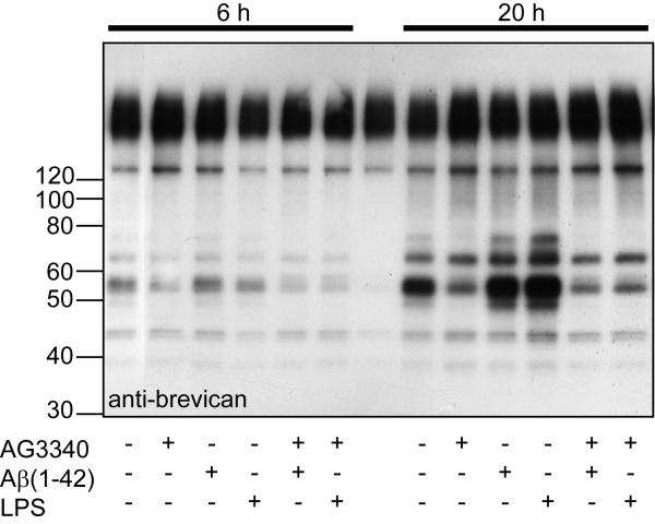
Aggregation of amyloid-beta (Abeta) in the forebrain of Alzheimer's disease (AD) subjects may disturb the molecular organization of the extracellular microenvironment that modulates neural and synaptic plasticity. Proteoglycans are major components of this extracellular environment. To test the hypothesis that Abeta, or another amyloid precursor protein (APP) dependent mechanism modifies the accumulation and/or turnover of extracellular proteoglycans, we examined whether the expression and processing of brevican, an abundant extracellular, chondroitin sulfate (CS)-bearing proteoglycan, were altered in brains of Abeta-depositing transgenic mice (APPsw - APP gene bearing the Swedish mutation) as a model of AD. The molecular size of CS chains attached to brevican was smaller in hippocampal tissue from APPsw mice bearing Abeta deposits compared to non-transgenic mice, likely because of changes in the CS chains. Also, the abundance of the major proteolytic fragment of brevican was markedly diminished in extracts from several telencephalic regions of APPsw mice compared to non-transgenic mice, yet these immunoreactive fragments appeared to accumulate adjacent to the plaque edge. These results suggest that Abeta or APP exert inhibitory effects on proteolytic cleavage mechanisms responsible for synthesis and turnover of proteoglycans. As proteoglycans stabilize synaptic structure and inhibit molecular plasticity, defective brevican processing observed in Abeta-bearing mice and potentially end-stage human AD, may contribute to deficient neural plasticity.


Figures

Figure 1
Figure 1
Detection of brevican isoforms and proteolytic degradation by endogenous proteases at specific cleavage sites. Brevican is secreted as a >145 kD protein bearing 1-3 CS chains (A). Brevican is also secreted as the holoprotein without CS chains at 145 kD (B). When probed on western blot with an N-terminal antibody (BD Biosciences, San Jose, CA) three immunoreactive bands appear;: a >145 smear (glycosylated brevican), the 145 kD core protein, and a ~55 kD proteolytic fragment (C, D, E and F). Arrows in (A) and (B) indicate proteolytic cleavage sites. Fragments of brevican are generated by endogenous proteases, the MMPs (D) and ADAMTSs (E). Each has a distinct, specific cleavage site sequence on the brevican protein. Shown here are the specific cleavage sequences for the MMPs and ADAMTSs in mouse, rat and human brevican (based on data from Nakamura et al. 2000). The MMP cleavage-site is 35 amino acid residues upstream from the ADAMTS-specific site (D and E). Distinct “neoepitope” antibodies recognize the MMP- and ADAMTS-derived cleavage fragments of brevican in mouse brain extracts subjected to Western blot on 4-20% gradient SDS-PAGE gels (F). Anti-SAHPSA recognizes the 53 kD, MMP-derived fragment of brevican (F; middle panel) whereas anti-EAMESE detects the 55 kD, ADAMTS-derived form (F; right panel). Mixing the two anti-bodies detects a “thicker” band in this region (F; left panel). “M” indicates molecular weight markers in (F). Antibodies recognize distinct products after proteolytic cleavage with hrADAMTS4 or hrMMP-2 (G). Proteoglycan purified from mouse brain was incubated with 50 nM hrADAMTS-4 (G, lane 1), 50 nM hrADAMTS4 + 5 mM EDTA (G, lane 2), 50 nM hrMMP-2 (G, lane 3) or 50 nM hrMMP-2 + 5 mM EDTA (G, lane 4) and immunoblotted for brevican. Note that the ADAMTS-derived brevican fragment was selectively recognized by anti-EAMESE and the brevican MMP product was recognized by anti-SAHPSA.
Figure 2
Figure 2
Immunoreactivity of Aβ and brevican isoforms in APPsw (+) transgenic mice compared to littermate non-transgenic (−) mice. Low power micrograph of immunoreactive Aβ burden in forebrain of 15-16 month old non-transgenic and APPsw mice (A). Hippocampal proteins from 15-16 month old non-transgenic (−)(n=3) and APPsw (+)(n=3) mice were separated on 4-20% SDS-PAGE, transferred to PVDF membrane and probed with anti-Aβ 95-2-5 (B). Note high and low molecular weight Aβ immunoreactivity (arrows). Lane “m” indicates molecular weight markers, lanes “h” are human frontal cortex tissue samples from patients diagnosed with AD, lanes Aβ 10 and Aβ 1 are synthetic Aβ(1-42) at 10 ng and 1 ng per lane respectively (a). Same blot was probed for anti-GAPDH immunoreactivity to normalize for protein loading (b). Identical hippocampal extracts were subjected to Western blot on 4-20% SDS-PAGE gels and probed for anti-brevican, anti-EAMESE and anti-SAHPSA (C). Densitometric semi-quantitative analysis of the western blots in (C) as expressed in arbitrary densitometric units (D). There was no change in abundance of >145 kD brevican protein in hippocampus, although an an identifiable molecular weight shift was apparent in APPsw extract. An increase in the abundance of the core 145 kD brevican (p ≤ 0.05) was accompanied by a decrease in the generalized N-terminal fragment of brevican in APPsw extracts (p ≤ 0.05). A marked decrease in the abundance of the brevican fragment generated by MMP-mediated proteolytic cleavage (p ≤ 0.05) was observed, denoted by anti-SAHPSA immunoreactivity, in hippocampal samples of APPsw mice.
Figure 3
Figure 3
Cha'se, N-glycanase and O-glycanase (+ sialidase) treatment of hippocampal extract derived from 15-16 month old non-transgenic and APPsw mice (A). Enzyme-treated-hippocampal proteins were separated on 6% Tris-glycine SDS-PAGE gels, blotted to PVDF and probed with an anti-brevican antibody. In samples from both genotypes, glycosidase treatments released the brevican core proteins to migrate to lower molecular weights indicating the presence N-and O-linked oligosaccharides and CS chains (A). The lower block of the image designated by the “star and brackets” indicates that the portion of the blot was exposed for a longer period of time compared to the upper region. (B) Hippocampal samples from three non-transgenic and three APPsw animals, with (+) and without (−) treatment with Ch'ase, subjected to SDS-PAGE, blotted, and probed with anti-brevican. Note the consistent migration difference in CS-bearing brevican, and the difference in the abundance of the core protein between the genotypes both before Ch'ase treatment but the little difference in abundance after Ch'ase treatment. Representative blots are shown from experiments that were repeated once.
Figure 4
Figure 4
Quantitative reverse transcriptase-polymerase chain reaction (qRT-PCR) for chondroition synthase synthesizing enzymes using RNA isolated from frontal cortex of non-transgenic (n = 6) and APPsw mice (n = 8). Threshold cycle number (2−Ct) for the gene of interest was divided by the threshold for the GAPDH gene (2−Ct) for each sample. There were no differences in expression for any of the five transcripts. Csgalnact1, Csgalnact2 (chondroitin sulfate N-acetyl-galactosaminyltransferase-1 and -2); Chsy1, Chsy2 (chondroitin sulfate synathase-1 and -2); Chpf (chon-droitin polymerizing factor).
Figure 5
Figure 5
Immunohistochemical localization of brevican (rb18 monoclonal), the proteolytically cleaved fragments of brevican (rabbit polyclonal against the ADAMTS-derived fragment, anti-EAMESE; MMP-derived fragment, anti-SAHPSA; and Aβ plaques (95-2-5 rabbit polyclonal) in APPsw transgenic mice (representative from n = 6 APPsw mice). Epifluorescent micrographs of brevican immunoreactivity (A, H, K), anti-EAMESE immunoreactivity (D, G), anti-SAHPSA immunoreactivity (J) and Aβ (B, E) in fixed frontal/parietal cortical sections. Merged composites (C, F, I & L). All images captured at 200x magnification.
Figure 6
Figure 6
Growth medium was changed to serum-free medium for 2 h and then cultured BV2 cells were treated with 2 μM Aβ(1-42) or 2 μg/ml LPS in the absence and the presence of the MMP inhibitor AG3340 (selective for MMP-2 and MMP-9). Two h later, 6 ug/well of a DEAE-purified preparation of proteoglycan was added for an additional 6 h (left panel) and 20 h (right panel). Supernatant was collected and subjected to Western blot for anti-brevican, anti-EAMESE and anti-SAHPSA.

Similar articles

Cited by

References

    1. Ajmo JM, Eakin AK, Hamel MG, Gottschall PE. Discordant localization of WFA reactivity and brevican/ADAMTS-derived fragment in rodent brain. BMC Neurosci. 2008;9:14. - PMC - PubMed
    1. Aldridge GM, Podrebarac DM, Greenough WT, Weiler IJ. The use of total protein stains as loading controls: an alternative to high-abundance single-protein controls in semi-quantitative immunoblotting. J Neurosci Methods. 2008;172:250–254. - PMC - PubMed
    1. Arendt T. Synaptic degeneration in Alzheimer's disease. Acta Neuropathol. 2009;118:167–179. - PubMed
    1. Asahina M, Yoshiyama Y, Hattori T. Expression of matrix metalloproteinase-9 and urinary-type plasminogen activator in Alzheimer's disease brain. Clin Neuropathol. 2001;20:60–63. - PubMed
    1. Backstrom JR, Lim GP, Cullen MJ, Tokes ZA. Matrix metalloproteinase-9 (MMP-9) is synthesized in neurons of the human hippocampus and is capable of degrading the amyloid-beta peptide (1-40) J Neurosci. 1996;16:7910–7919. - PMC - PubMed

Publication types

MeSH terms