. 2010 Mar;75(6):1529-38.
doi: 10.1111/j.1365-2958.2010.07076.x. Epub 2010 Feb 17.

The IsdG-family of haem oxygenases degrades haem to a novel chromophore

Affiliations

The IsdG-family of haem oxygenases degrades haem to a novel chromophore

Michelle L Reniere et al. Mol Microbiol. 2010 Mar.

Abstract

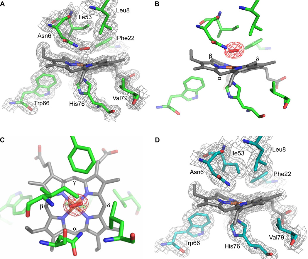
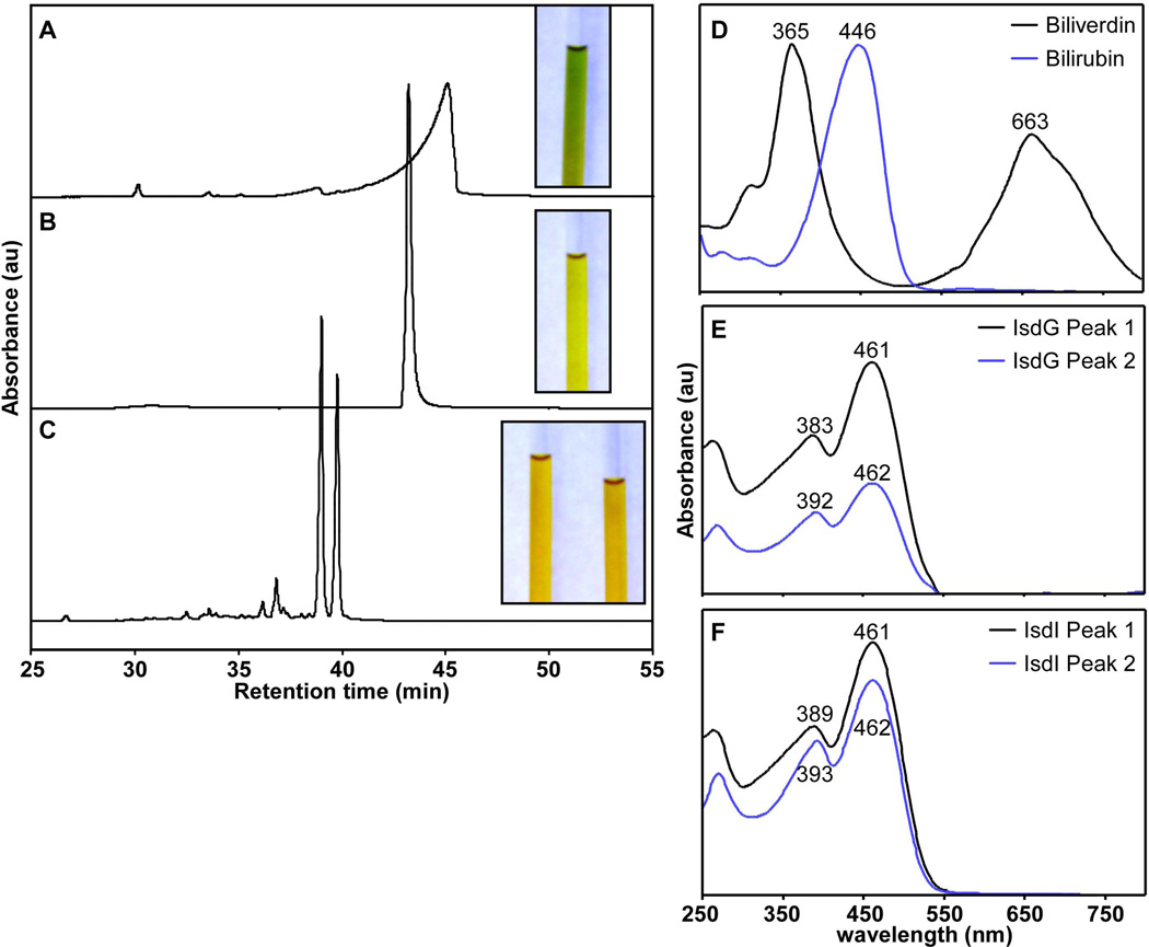
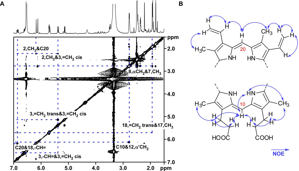
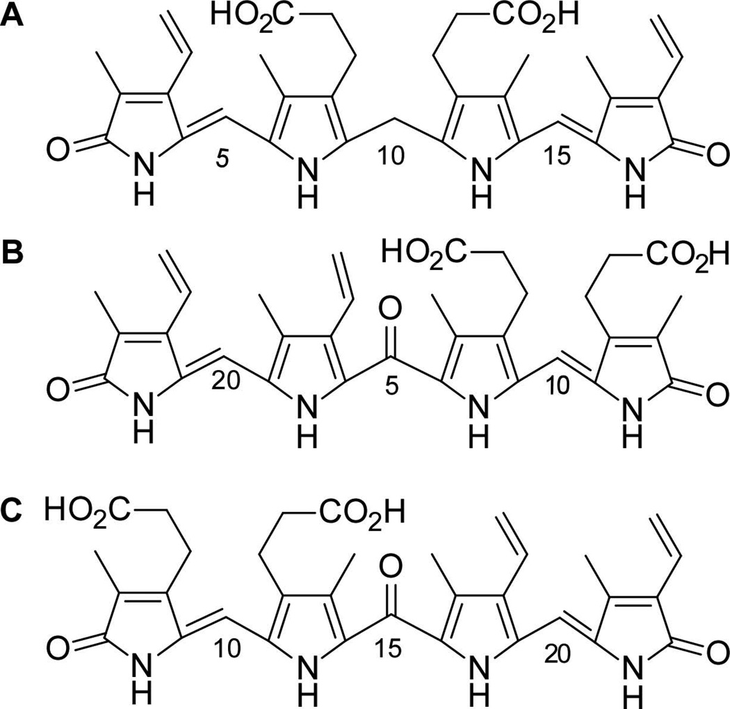
Enzymatic haem catabolism by haem oxygenases is conserved from bacteria to humans and proceeds through a common mechanism leading to the formation of iron, carbon monoxide and biliverdin. The first members of a novel class of haem oxygenases were recently identified in Staphylococcus aureus (IsdG and IsdI) and were termed the IsdG-family of haem oxygenases. Enzymes of the IsdG-family form tertiary structures distinct from those of the canonical haem oxygenase family, suggesting that IsdG-family members degrade haem via a unique reaction mechanism. Herein we report that the IsdG-family of haem oxygenases degrade haem to the oxo-bilirubin chromophore staphylobilin. We also present the crystal structure of haem-bound IsdI in which haem ruffling and constrained binding of oxygen is consistent with cleavage of the porphyrin ring at the beta- or delta-meso carbons. Combined, these data establish that the IsdG-family of haem oxygenases degrades haem to a novel chromophore distinct from biliverdin.


Figures

Fig. 1
Fig. 1
Crystal structure of the active site of IsdI-heme. The structure of IsdI (green) bound to heme (gray) reveals density on the distal side modeled as a dioxygen species (red) liganded to the heme-iron. A. 2Fo - Fc map contoured at 1σ. B. A Fo - Fc omit map for oxygen is contoured at 3σ. The heme α-, β-, and δ-meso carbons are labeled. The γ-meso carbon is buried in the protein. C. The same figure as in b, but looking down perpendicular to the heme from the distal side. The heme α-, β-, γ-, and δ-meso carbons are labeled. D. Crystals of IsdI (cyan) bound to heme (gray) were reduced for 10 minutes in 50 mM (excess) dithionite. The 2Fo - Fc map (light gray) is contoured at 1σ. Heme and selected amino acids are depicted in sticks and are labeled. Nitrogen, oxygen and iron atoms are colored blue, red, and orange, respectively.
Fig. 2
Fig. 2
Purification and optical spectra of heme degradation products. HPLC tracings of heme degradation products separated on a linear acetonitrile gradient. Insets are images of the heme degradation products dissolved in DMSO. The contrast of the photos was adjusted to present the clearest images. A. Biliverdin purification monitored at 405 nm. B. Bilirubin purification monitored at 405 nm. C. The product of IsdI-catalyzed heme degradation monitored at 465 nm. The two prominent peaks were collected separately and both product images are shown (inset). IsdG- and IsdI-catalyzed heme degradation products have overlapping spectra so only that of IsdI is shown for simplicity. D–F. Optical spectra of each fraction as measured by the photodiode array detector upon elution.
Fig. 3
Fig. 3
NOESY correlations of IsdG product peak 1. A. NOESY spectrum (in d6-DMSO) of the first eluting product focused on key regions that demonstrate correlations between the α- and γ-bridge CHs and neighboring protons. The second eluting product from the IsdG-catalyzed reaction and the IsdI-catalyzed products have similar NOESY spectra, and are shown in Fig. S5–S7. B. Two fragments of a bilirubin-like molecule with NOE correlations (arrows). Carbon 10 and 20 are labeled according to standard bilirubin nomenclature in which C10 corresponds to the γ-meso carbon and C20 corresponds to the α-meso carbon of the porphyrin ring.
Fig. 4
Fig. 4
Linear representations of heme degradation products. A. Bilirubin. B. 5-oxo-δ-bilirubin, the first eluting product. C. 15-oxo-β-bilirubin, the second product to elute upon HPLC purification. The bilirubin carbon numbering scheme was used for the IsdG and IsdI products for simplicity.
Fig. 5
Fig. 5
Tandem LC- HRESIMS. A. ESI-MS/MS spectrum of fragment ions selecting for 599.3 m/z for 5-oxo-δ-bilirubin. B. ESI-MS/MS spectrum of fragment ions selecting for 599.3 m/z for 15-oxo-β-bilirubin. C,D. Fragmentation scheme corresponding to the masses and molecular formulas obtained in A and B respectively. ESI-MS/MS spectra for IsdI-catalyzed products are similar.

References

    1. Bullen JJ, Griffiths E. Iron and Infection: Molecular, Physiological and Clinical Aspects. New York: John Wiley and Sons; 1999.
    1. Chen Q, Huggins MT, Lightner DA, Norona W, McDonagh AF. Synthesis of a 10-Oxo-Bilirubin: Effects of the Oxo Group on Conformation, Transhepatic Transport, and Glucuronidation. Journal of the American Chemical Society. 1999;121:9253–9264.
    1. Chim N, Iniguez A, Nguyen TQ, Goulding CW. Unusual Diheme Conformation of the Heme-Degrading Protein from Mycobacterium tuberculosis. J Mol Biol. 2009 - PMC - PubMed
    1. Chung SW, Hall SR, Perrella MA. Role of haem oxygenase-1 in microbial host defence. Cell Microbiol. 2009;11:199–207. - PMC - PubMed
    1. Cole WJ, Chapman DJ, Siegelman HW. The structure and properties of phycocyanobilin and related bilatrienes. Biochemistry. 1968;7:2929–2935. - PubMed

Publication types

MeSH terms

LinkOut - more resources