

Human interphase chromosomes: a review of available molecular cytogenetic technologies
- PMID: 20180947
- PMCID: PMC2830939
- DOI: 10.1186/1755-8166-3-1
Human interphase chromosomes: a review of available molecular cytogenetic technologies
Abstract
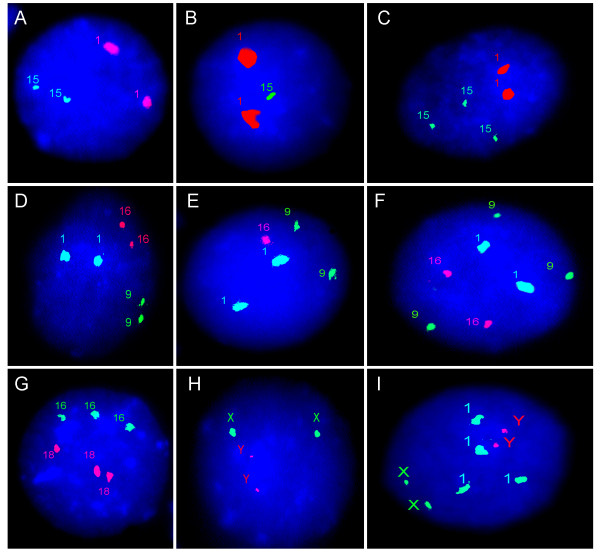
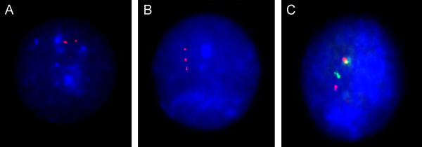
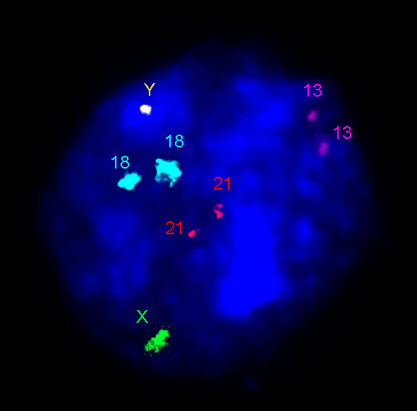
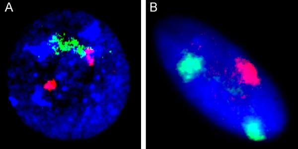
Human karyotype is usually studied by classical cytogenetic (banding) techniques. To perform it, one has to obtain metaphase chromosomes of mitotic cells. This leads to the impossibility of analyzing all the cell types, to moderate cell scoring, and to the extrapolation of cytogenetic data retrieved from a couple of tens of mitotic cells to the whole organism, suggesting that all the remaining cells possess these genomes. However, this is far from being the case inasmuch as chromosome abnormalities can occur in any cell along ontogeny. Since somatic cells of eukaryotes are more likely to be in interphase, the solution of the problem concerning studying postmitotic cells and larger cell populations is interphase cytogenetics, which has become more or less applicable for specific biomedical tasks due to achievements in molecular cytogenetics (i.e. developments of fluorescence in situ hybridization -- FISH, and multicolor banding -- MCB). Numerous interphase molecular cytogenetic approaches are restricted to studying specific genomic loci (regions) being, however, useful for identification of chromosome abnormalities (aneuploidy, polyploidy, deletions, inversions, duplications, translocations). Moreover, these techniques are the unique possibility to establish biological role and patterns of nuclear genome organization at suprachromosomal level in a given cell. Here, it is to note that this issue is incompletely worked out due to technical limitations. Nonetheless, a number of state-of-the-art molecular cytogenetic techniques (i.e multicolor interphase FISH or interpahase chromosome-specific MCB) allow visualization of interphase chromosomes in their integrity at molecular resolutions. Thus, regardless numerous difficulties encountered during studying human interphase chromosomes, molecular cytogenetics does provide for high-resolution single-cell analysis of genome organization, structure and behavior at all stages of cell cycle.
Figures
Similar articles
-
Visualization of interphase chromosomes in postmitotic cells of the human brain by multicolour banding (MCB).Chromosome Res. 2006;14(3):223-9. doi: 10.1007/s10577-006-1037-6. Epub 2006 Apr 20. Chromosome Res. 2006. PMID: 16628493
-
[Molecular cytogenetic methods for studying interphase chromosomes in human brain cells].Genetika. 2010 Sep;46(9):1171-4. Genetika. 2010. PMID: 21061610 Russian.
-
The DNA-based structure of human chromosome 5 in interphase.Am J Hum Genet. 2002 Nov;71(5):1051-9. doi: 10.1086/344286. Epub 2002 Oct 7. Am J Hum Genet. 2002. PMID: 12370837 Free PMC article.
-
Cytogenetic and molecular cytogenetic analysis of B cell chronic lymphocytic leukemia: specific chromosome aberrations identify prognostic subgroups of patients and point to loci of candidate genes.Leukemia. 1997 Apr;11 Suppl 2:S19-24. Leukemia. 1997. PMID: 9178833 Review.
-
[Molecular cytogenetic techniques and their application in clinical diagnosis].Med Wieku Rozwoj. 2004 Jan-Mar;8(1):7-24. Med Wieku Rozwoj. 2004. PMID: 15557693 Review. Polish.
Cited by
-
Single cell genomics of the brain: focus on neuronal diversity and neuropsychiatric diseases.Curr Genomics. 2012 Sep;13(6):477-88. doi: 10.2174/138920212802510439. Curr Genomics. 2012. PMID: 23449087 Free PMC article.
-
Ontogenetic and Pathogenetic Views on Somatic Chromosomal Mosaicism.Genes (Basel). 2019 May 19;10(5):379. doi: 10.3390/genes10050379. Genes (Basel). 2019. PMID: 31109140 Free PMC article. Review.
-
Molecular cytogenetic diagnosis and somatic genome variations.Curr Genomics. 2010 Sep;11(6):440-6. doi: 10.2174/138920210793176010. Curr Genomics. 2010. PMID: 21358989 Free PMC article.
-
Challenges in the clinical advancement of cell therapies for Parkinson's disease.Nat Biomed Eng. 2023 Apr;7(4):370-386. doi: 10.1038/s41551-022-00987-y. Epub 2023 Jan 12. Nat Biomed Eng. 2023. PMID: 36635420 Free PMC article. Review.
-
Diversity of sex chromosome abnormalities in a cohort of 95 Indonesian patients with monosomy X.Mol Cytogenet. 2011 Oct 12;4:23. doi: 10.1186/1755-8166-4-23. Mol Cytogenet. 2011. PMID: 21992692 Free PMC article.
References
-
- Gersen SL, Keagle MB. The principles of clinical cytogenetics. 2. Totowa, NJ: Humana Press; 2005.
-
- Liehr T, Claussen U. Multicolor-FISH approaches for the characterization of human chromosomes in clinical genetics and tumor cytogenetics. Curr Genomics. 2002;3:231–235. doi: 10.2174/1389202023350525. - DOI
-
- Liehr T, Starke H, Weise A, Lehrer H, Claussen U. Multicolor FISH probe sets and their applications. Histol Histopathol. 2004;19:229–237. - PubMed
LinkOut - more resources
Full Text Sources
