

Benign myoepithelioma of the lung - a case report and review of the literature
- PMID: 20180958
- PMCID: PMC2828429
- DOI: 10.1186/1757-1626-3-25
Benign myoepithelioma of the lung - a case report and review of the literature
Abstract
Introduction: Benign myoepithelioma is extremely rare in the lung, to the best of our knowledge; only five cases have been reported in the literature.
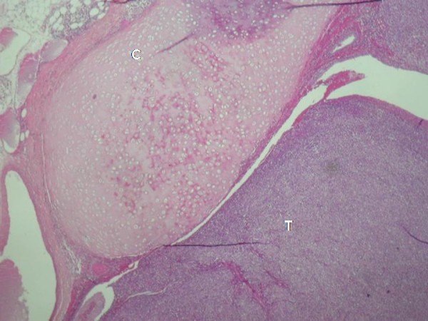
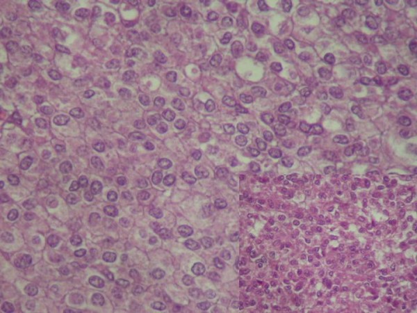
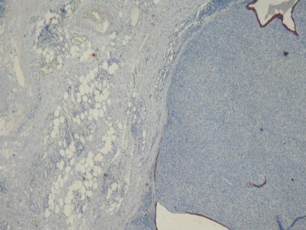
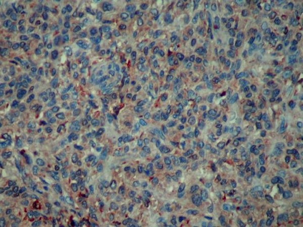
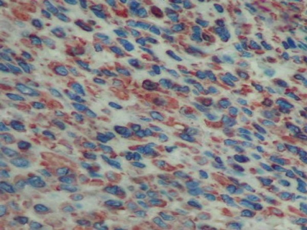
Case report: An 18-years woman complained from tiredness and fever during four months. Laboratory findings and fibroscopies were normal. CT of the thorax demonstrated a nodule in the left segment of the Fowler. Left inferior lobectomy was performed comporting a firm nodule of 25 mm, lifting the bronchial mucous membrane. Histologically, there was a proliferation of small cells of a plasmocytoid-type, with a predominantly whorled pattern. No mitotic activity or necrosis was seen in the tumor. Immuhistochemically, the tumor cells positive for smooth muscle actin, vimentine, and S100 protein. They were negatives for cytokeratine, chromogranine and HMB45. The diagnosis of benign myoepithelioma of the lung is so confirmed. The patient recovered well at 6 months follow-up.
Conclusion: Benign myoepithelioma is a rare pulmonary neoplasm distinct from pleomorphic adenoma, which should be considered in the differential diagnosis of lung nodules.
Figures
Similar articles
-
Benign myoepithelioma of the lung: a case report and review of the literature.Arch Pathol Lab Med. 2001 Nov;125(11):1494-6. doi: 10.5858/2001-125-1494-BMOTL. Arch Pathol Lab Med. 2001. PMID: 11698012 Review.
-
Plasmacytoid myoepithelioma of the palate in a child.Int J Paediatr Dent. 2007 May;17(3):223-7. doi: 10.1111/j.1365-263X.2006.00785.x. Int J Paediatr Dent. 2007. PMID: 17397468
-
Plasmacytoid myoepithelioma of the palate. Report of one case and review of the literature.Med Oral Patol Oral Cir Bucal. 2007 Dec 1;12(8):E552-5. Med Oral Patol Oral Cir Bucal. 2007. PMID: 18059237 Review.
-
Cutaneous myoepithelioma in the foot: case report.Foot Ankle Spec. 2013 Jun;6(3):239-41. doi: 10.1177/1938640012470713. Epub 2012 Dec 21. Foot Ankle Spec. 2013. PMID: 23263678
-
What is hiding behind S100 protein and SOX10 positive oncocytomas? Oncocytic pleomorphic adenoma and myoepithelioma with novel gene fusions in a subset of cases.Hum Pathol. 2020 Sep;103:52-62. doi: 10.1016/j.humpath.2020.07.009. Epub 2020 Jul 13. Hum Pathol. 2020. PMID: 32673681
Cited by
-
A large mediastinal benign myoepithelioma effacing the entire hemithorax: case report with literature review.Diagn Pathol. 2015 Jul 14;10:100. doi: 10.1186/s13000-015-0340-y. Diagn Pathol. 2015. PMID: 26170201 Free PMC article. Review.
-
Tracheal myoepithelioma resected by using rigid bronchoscopy: a case report and review of the literature.J Cardiothorac Surg. 2022 May 23;17(1):125. doi: 10.1186/s13019-022-01880-0. J Cardiothorac Surg. 2022. PMID: 35606819 Free PMC article. Review.
-
Thoracic Myoepithelial Tumors: A Pathologic and Molecular Study of 8 Cases With Review of the Literature.Am J Surg Pathol. 2016 Feb;40(2):212-23. doi: 10.1097/PAS.0000000000000560. Am J Surg Pathol. 2016. PMID: 26645726 Free PMC article.
References
-
- Colby TV, Koss MN, Travis WD. In: Tumors of the lower respiratory tract. Rosai J, editor. Washington, DC: Armed Forces Institute of Pathology; 1995.
-
- Strickler JG, Hegstrom J, Thomas MJ, Yousem SA. Myoepithelioma of the lung. Arch Pathol Lab Med. 1987;111:1082–5. - PubMed
-
- Veeramachaneni R, Gulick J, Halldosson AO, Van TT, Zhang PL, Herrera GA. Benign Myoepithelioma of the lung. A case report and review of the literature. Arch Pathol Lab Med. 2001;125:1494–6. - PubMed
-
- El Mezni F, Zeddini A, Hamzaoui A, Ismail O, Ghrairi H, Ben Miled K, Smati B, Kilani T. Myoépithéliome benin du poumon. Rev Pneumol Clin. 2004;60(5):282–284. - PubMed
LinkOut - more resources
Full Text Sources
