Comparative Study
. 2010 Jan 26:5:9.
doi: 10.1186/1746-1596-5-9.

Utility of immunohistochemical markers in differentiating benign from malignant follicular-derived thyroid nodules

Affiliations
Comparative Study

Utility of immunohistochemical markers in differentiating benign from malignant follicular-derived thyroid nodules

Husain A Saleh et al. Diagn Pathol. .

Abstract

Background: Thyroid nodules are common among adults though only a small percentage is malignant, which can histologically mimic benign nodules. Accurate diagnosis of these thyroid nodules is critical for the proper clinical management.

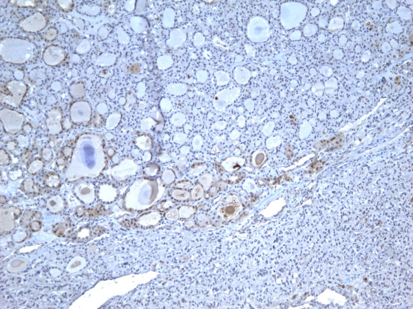
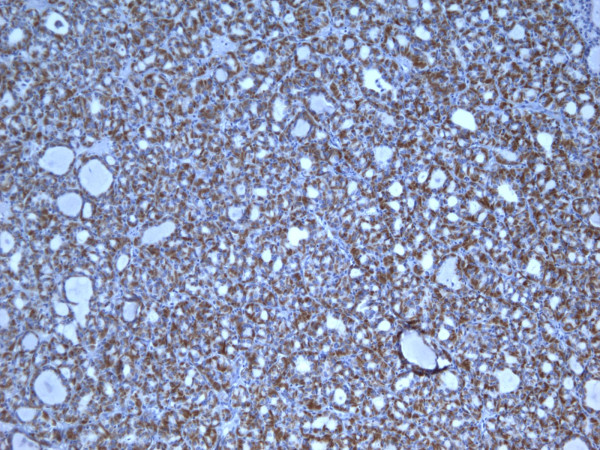
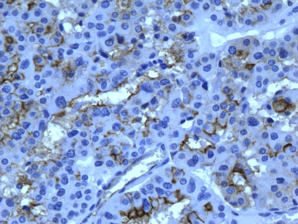
Methods: We investigated immunoexpression in 98 surgically removed benign thyroid nodules including 52 hyperplastic nodules (HN) and 46 follicular/Hurthle cell adenomas (FA), and 54 malignant tumors including 22 follicular carcinoma (FC), 20 classic papillary carcinoma (PTC), and 12 follicular variant papillary carcinoma (FVPC).

Results: The staining results showed that malignant tumors express galectin-3, HBME-1, CK19 and Ret oncoprotein significantly more than benign nodules. The sensitivity of these markers for the distinction between benign and malignant lesions ranged from 83.3% to 87%. The sensitivity of two-marker panels was not significantly different. Immunoexpression was usually diffuse and strong in malignant tumors, and focal and weak in the benign lesions.

Conclusion: Our findings indicate that these immunomarkers are significantly more expressed in malignant tumors compared to benign lesions and may be of additional diagnostic value when combined with routine histology.


Figures

Figure 1
Figure 1
Immunohistochemical stain of hyperplastic colloid nodule shows the cells are negative for galectin-3 (100×).
Figure 2
Figure 2
Immunohistochemical stain of hyperplastic colloid nodule shows the cells are negative for Ret oncoprotein (100×).
Figure 3
Figure 3
Immunohistochemical stain of hyperplastic colloid nodule shows the cells are negative, for HBME-1 (100×).
Figure 4
Figure 4
Immunohistochemical stain of hyperplastic colloid nodule shows the cells are negative for CK19 (100×).
Figure 5
Figure 5
Immunohistochemical stain of follicular adenoma shows the tumor cells focally reactive for galectin-3 (100×).
Figure 6
Figure 6
Immunohistochemical stain of follicular adenoma shows the tumor cells focally reactive for Ret oncoprotein (100×).
Figure 7
Figure 7
Immunohistochemical stain of follicular adenoma shows the tumor cells focally reactive for HBME-1 (100×).
Figure 8
Figure 8
Immunohistochemical stain of follicular adenoma shows the tumor cells focally reactive CK19 (400×).
Figure 9
Figure 9
Immunohistochemical stain reveals that tumor cells in follicular carcinoma are strongly and diffusely positive for galectin-3 (400×).
Figure 10
Figure 10
Immunohistochemical stain reveals that tumor cells in follicular carcinoma are strongly and diffusely positive for Ret oncoprotein (400×).
Figure 11
Figure 11
Immunohistochemical stain reveals that tumor cells in follicular carcinoma are strongly and diffusely positive for HBME-1 (400×).
Figure 12
Figure 12
Immunohistochemical stain reveals that tumor cells in follicular carcinoma are strongly and diffusely positive for CK19 (400×).
Figure 13
Figure 13
Immunohistochemical stain reveals that the tumor cells of classic papillary thyroid carcinoma are strongly and diffusely positive for galectin-3 (100×).
Figure 14
Figure 14
Immunohistochemical stain reveals that the tumor cells of classic papillary thyroid carcinoma are strongly and diffusely positive for Ret oncoprotein (100×).
Figure 15
Figure 15
Immunohistochemical stain reveals that the tumor cells of classic papillary thyroid carcinoma are strongly and diffusely positive for HBME-1 (100×).
Figure 16
Figure 16
Immunohistochemical stain reveals that the tumor cells of classic papillary thyroid carcinoma are strongly and diffusely positive for CK19 (100×).
Figure 17
Figure 17
Immunohistochemical stain shows that the tumor cells of follicular variant of papillary carcinoma are strongly and diffusely positive for galectin-3 (100×).
Figure 18
Figure 18
Immunohistochemical stain shows that the tumor cells of follicular variant of papillary carcinoma are strongly and diffusely positive for Ret oncoprotein (100×).
Figure 19
Figure 19
Immunohistochemical stain shows that the tumor cells of follicular variant of papillary carcinoma are strongly and diffusely positive for HBME-1 (400×).
Figure 20
Figure 20
Immunohistochemical stain shows that the tumor cells of follicular variant of papillary carcinoma are strongly and diffusely positive for CK19 (400×).

Similar articles

Cited by

References

    1. Oestreicher-Kedem Y, Halpern M, Roizman P, Hardy B, Sulkes J, Feinmesser R, Stern Y. Diagnostic value of galectin-3 as a marker for malignancy in follicular patterned thyroid lesions. Head Neck. 2004;26(11):960–6. doi: 10.1002/hed.20087. - DOI - PubMed
    1. Papotti M, Rodriguez J, De Pompa R, Bartolazzi A, Rosai J. Galectin-3 and HBME-1 expression in well-differentiated thyroid tumors with follicular architecture of uncertain malignant potential. Mod Pathol. 2005;18(4):541–6. doi: 10.1038/modpathol.3800321. - DOI - PubMed
    1. Prasad ML, Pellegata NS, Huang Y, Nagaraja HN, de la Chapelle A, Kloos RT. Galectin-3, fibronectin-1, CITED-1, HBME1 and cytokeratin-19 immunohistochemistry is useful for the differential diagnosis of thyroid tumors. Mod Pathol. 2005;18(1):48–57. doi: 10.1038/modpathol.3800235. - DOI - PubMed
    1. Rosai J. Papillary thyroid carcinoma: a root-and-branch rethink. Am J Clin Pathol. 2008;130(5):683–6. doi: 10.1309/AJCPBF63BWMCYSLW. - DOI - PubMed
    1. Sahoo S, Hoda SA, Rosai J, DeLellis RA. Cytokeratin 19 immunoreactivity in the diagnosis of papillary thyroid carcinoma: a note of caution. Am J Clin Pathol. 2001;116(5):696–702. doi: 10.1309/6D9D-7JCM-X4T5-NNJY. - DOI - PubMed

MeSH terms

Supplementary concepts

LinkOut - more resources