

Chlamydia trachomatis growth inhibition and restoration of LDL-receptor level in HepG2 cells treated with mevastatin
- PMID: 20181044
- PMCID: PMC2835644
- DOI: 10.1186/1476-5926-9-3
Chlamydia trachomatis growth inhibition and restoration of LDL-receptor level in HepG2 cells treated with mevastatin
Abstract
Background: Perihepatitis is rare but consistently occurring extragenital manifestation of untreated Chlamydia trachomatis infection. Despite of possible liver involvement in generalized C. trachomatis infection, the ability of the pathogen to propagate in the hepatic cells and its impact on liver functions is not thoroughly investigated. The effect of mevastatin, an inhibitor of 3-hydroxy-3-methylglutaryl CoA reductase, on C. trachomatis growth in human hepatoma cell line HepG2 has been studied. Bacterial growth was assessed by immunostaining with FITC-labeled monoclonal antibody against chlamydial lipopolysaccharide and by RT-PCR for two chlamydial genetic markers (16S rRNA and euo).
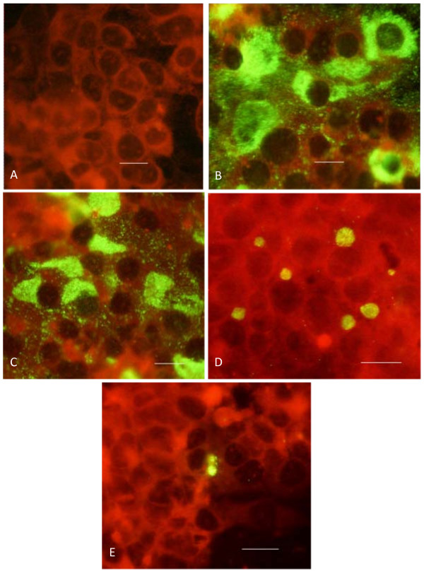
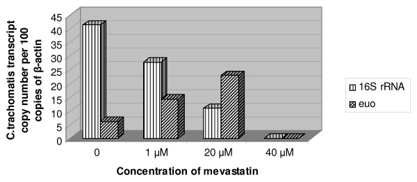
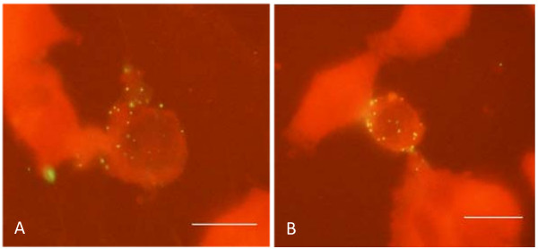
Results: Chlamydial inclusion bodies were seen in approximately 50% of hepatocytes at 48 hours in the post infection period. Lysates obtained from infected hepatocytes were positive in the infective progeny test at 48 and especially in 72 hours after infection initiation. It has been shown that chlamydial infection in hepatocytes also leads to the decline of LDL-receptor mRNA which reflects infection multiplicity rate. Additions of mevastatin (1, 20 and 40 microM) 1 hour before inoculation restored and upregulated LDL-receptor mRNA level in a dose-dependent manner. Mevastatin treatment had no effect on internalization of chlamydial particles. However it reduced drastically the number of chlamydial 16S rRNA and euo transcripts as well as overall infection rate in HepG-2 cells. Complete eradication of infection has been seen by immunofluorescent staining at 40 microM mevastatin concentration, when expression level of chlamydial 16S rRNA and euo was undetectable. Lower concentration of mevastatin (20 microM) promoted euo expression level and the appearance of atypically small chlamydial inclusions, while there was a noticeable reduction in the number of infected cells and 16S rRNA transcripts.
Conclusions: C. trachomatis can efficiently propagate in hepatocytes affecting transcription rate of some liver-specific genes. Ongoing cholesterol synthesis is essential for chlamydial growth in hepatocytes. Inhibitors of cholesterol biosynthesis can supplement conventional strategy in the management of C. trachomatis infection.
Figures
References
-
- Yang JL, Hong KC, Schachter J, Moncada J, Lekew T, House JI, Zhou Z, Neuwelt MD, Rutar T, Halfpenny C, Shah N, Whitcher JP, Lietman TM. Detection of Chlamydia trachomatis ocular infection in trachoma-endemic communities by rRNA amplification. Invest Ophthalmol Vis Sci. 2009;50:90–94. doi: 10.1167/iovs.08-2247. - DOI - PubMed
-
- Bilenki L, Wang S, Yang J, Fan Y, Joyee AG, Yang X. Chlamydia trachomatis NK T cell activation promotes infection in vivo. J Immunol. 2005;175:3197–3206. - PubMed
-
- Roubalova K. A simultaneous detection of Chlamydia pneumoniae and Chlamydia trachomatis DNA by real-time PCR. Epidemiol Mikrobiol Imunol. 2007;56:166–173. - PubMed
LinkOut - more resources
Full Text Sources
Other Literature Sources