. 2010 Feb 2:7:3.
doi: 10.1186/1742-4933-7-3.

Differential impact of ageing on cellular and humoral immunity to a persistent murine gamma-herpesvirus

Affiliations

Differential impact of ageing on cellular and humoral immunity to a persistent murine gamma-herpesvirus

Eric J Yager et al. Immun Ageing. .

Abstract

Background: Oncogenic gamma-herpesviruses establish life-long infections in their hosts and control of these latent infections is dependent on continual immune surveillance. Immune function declines with age, raising the possibility that immune control of gamma-herpesvirus infection becomes compromised with increasing age, allowing viral reactivation and/or increased latent load, both of which are associated with the development of malignancies.

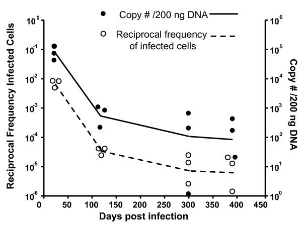
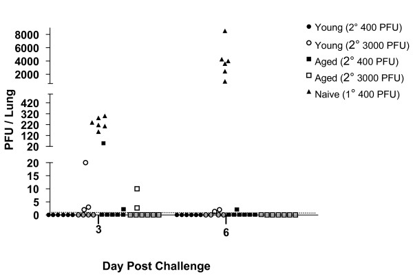
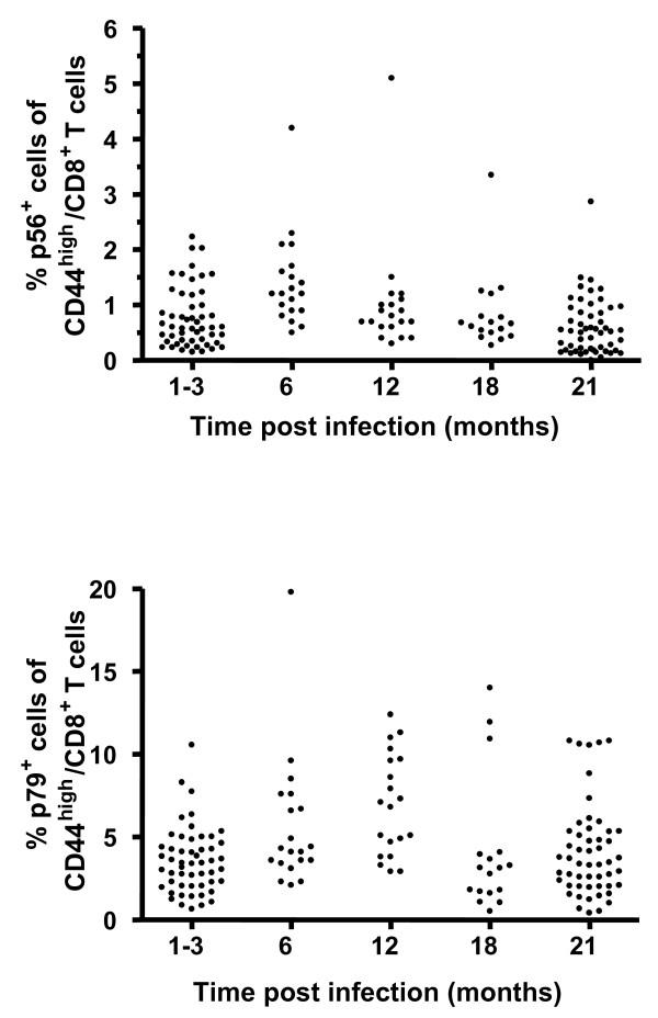
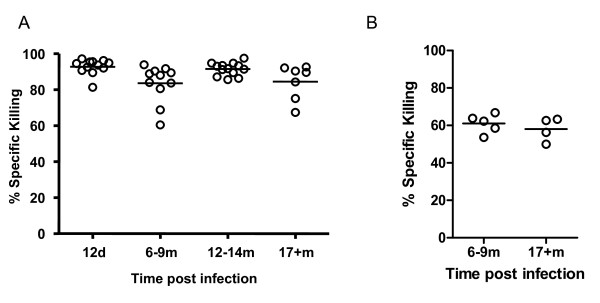
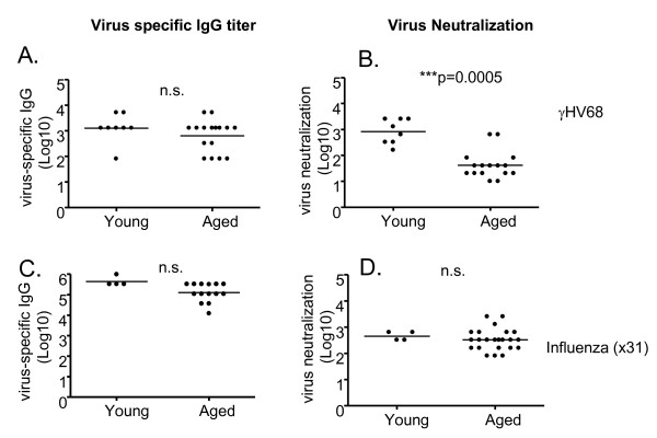
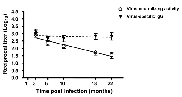
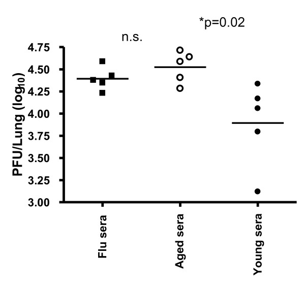
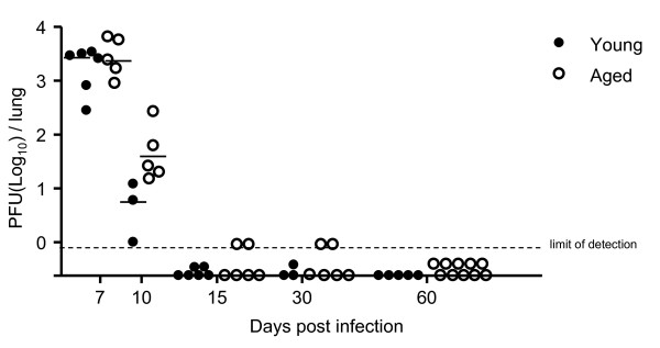
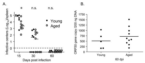
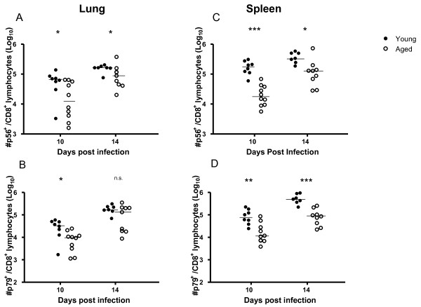
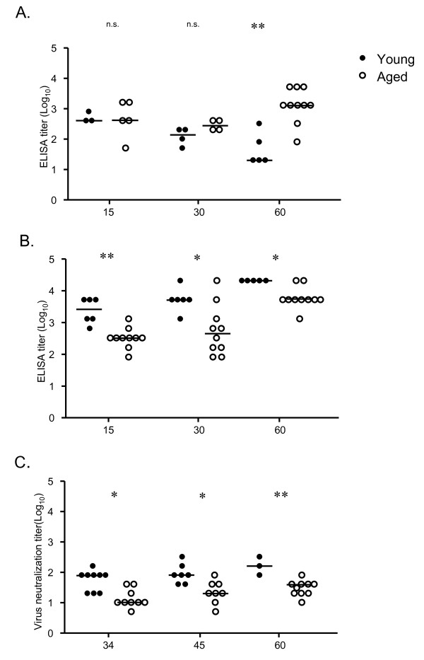
Results: In this study, we use the experimental mouse gamma-herpesvirus model, gammaHV68, to investigate viral immunity in aged mice. We found no evidence of viral recrudescence or increased latent load in aged latently-infected mice, suggesting that effective immune control of gamma-herpesvirus infection remains intact with ageing. As both cellular and humoral immunity have been implicated in host control of gammaHV68 latency, we independently examined the impact of ageing on gammaHV68-specific CD8 T cell function and antibody responses. Virus-specific CD8 T cell numbers and cytolytic function were not profoundly diminished with age. In contrast, whereas ELISA titers of virus-specific IgG were maintained over time, there was a progressive decline in neutralizing activity. In addition, although aged mice were able to control de novo acute infection with only slightly delayed viral clearance, serum titers of neutralizing antibody were reduced in aged mice as compared to young mice.

Conclusion: Although there is no obvious loss of immune control of latent virus, these data indicate that ageing has differential impacts on anti-viral cellular and humoral immune protection during persistent gammaHV68 infection. This observation has potential relevance for understanding gamma-herpesvirus immune control during disease-associated or therapeutic immunosuppression.


Figures

Figure 1
Figure 1
γHV68 latency in the spleen remains stable with age. The reciprocal frequency of latently-infected cells as determined by LDA/PCR is plotted on the left axis (open circles) and the genome copy number per 200 ng DNA is plotted on the right axis (closed circles) over time after intranasal infection with 400 PFU γHV68. The lines join the means of three individual mice at each timepoint.
Figure 2
Figure 2
Latently-infected aged mice are protected against re-infection by γHV68. Young mice (3 months pi; circles) and aged mice (19 months pi; squares) were intranasally challenged with γHV68 (400 PFU, closed symbols or 3000 PFU, open symbols). Naïve mice were challenged as a control (triangles). Symbols indicate the titers of lytic γHV68 virus detected in the lungs of individual mice at 3 and 6 dpi. The dotted line represents the limit of detection for the plaque assay used to measure viral titers. Chi-square analysis comparing numbers of mice with or without plaques detectable in lungs revealed significant differences (P < 0.05) only between the naïve mice challenged as a control and the other four groups.
Figure 3
Figure 3
Virus-specific CD8 T cell numbers are maintained with age. Mice were bled at various times following γHV68 infection and virus specific CD8 T cells were detected using MHC class I tetramer staining and flow cytometry. Symbols represent the frequency of ORF6487-495 (p56, upper panel) and ORF61524-531 (p79, lower panel) specific cells among CD44high CD8+ T cells found in the peripheral blood of individual mice at the indicated times post infection.
Figure 4
Figure 4
Virus-specific CD8 T cells present in latently-infected mice retain CTL function with age. Cytolytic activity of ORF61-specific CD8 T cells was measured at the indicated times post infection using a 16-17 h (Panel A) or a 4 h (Panel B) in vivo CTL assay as described in the Materials and Methods. Symbols represent specific killing calculated for individual mice. Bars indicate the medians calculated from data compiled from three independent experiments. Significance was assessed using the Mann-Whitney rank test (two-tailed, 95% confidence).
Figure 5
Figure 5
Serum titers of γHV68-specific antibody are maintained in latently infected mice with age, but serum neutralization activity wanes. The titers of virus-specific IgG were measured in the serum of latently-infected young (1-3 months p.i.) and aged (18-22 months p.i.) by ELISA (Panel A). Neutralization activity (measured in the same sera samples as A) were determined using an in vitro neutralization assay (Panel B). As a control, antibody titers (C) and neutralization activity (D) were measured in serum taken from mice (young and aged, as above) previously infected with influenza virus ×31. Symbols represent serum antibody titers and neutralization activities measured for individual mice. Bars indicate the medians calculated from the data shown. Significance was assessed using the Mann-Whitney rank test (two-tailed, 95% confidence). Ns; not significant.
Figure 6
Figure 6
Neutralizing titers of γHV68 decline progressively with time after infection. Serum titers of γHV68-specific IgG (closed triangles) and neutralizing antibody (open circles) were measured in individual mice at the indicated months post infection. Symbols represent the mean reciprocal titers (log10), ± standard deviation, calculated at each timepoint (≥ 8 mice analyzed per timepoint). A linear regression was performed analyzing time versus reciprocal titer for virus-specific IgG or neutralizing antibody. The slope for virus specific IgG did not differ significantly from zero (p = 0.6780), however the slope for virus neutralizing antibody did (p = 0.0085).
Figure 7
Figure 7
Sera from aged mice is less protective in vivo against acute γHV68 infection. One hundred microliters of sera from young (3 months p. i.) or aged (22 months p. i.) γHV68-infected animals was injected intravenously into naïve C57BL/6 mice one day prior to intranasal γHV68 infection (400 PFU). Control mice received convalescent sera from influenza virus-infected mice. Symbols represent lung viral titers measured in individual mice at 5 days post infection using a standard plaque assay. Bars indicate the medians calculated from the data shown. Significance was assessed using the Mann-Whitney rank test (two-tailed, 95% confidence). Ns; not significant
Figure 8
Figure 8
Aged mice clear lytic virus from their lungs following de novo γHV68 infection with only slightly delayed kinetics. The titers of lytic virus in the lungs of individual young (3 months old, closed symbols) and aged (19-24 months old, open symbols) C57BL/6 mice at the indicated days following de novo intranasal γHV68 infection (400 PFU) were measured using a standard plaque assay. The dotted line indicates the plaque assay's limit of detection.
Figure 9
Figure 9
Aged mice are capable of maintaining control over latent virus following de novo γHV68 infection. The levels of latent virus present in the spleens of γHV68-infected young (3 months old, closed symbols) and aged (19-24 months old, open symbols) mice at the indicated days post infection were measured using an infective center assay (Panel A) and a genome copy assay (Panel B), as described in the Material and Methods. The dotted line indicates the limit of detection of the infective center assay. Each symbol represents data obtained from an individual mouse and bars indicate the means calculated from the data shown.*; P ≤ 0.05 as determined using the Student's t-test. n.s.; not statistically significant.
Figure 10
Figure 10
Virus-specific CD8 T cells are generated in aged mice following de novo γHV68 infection. MHC class I tetramer staining and flow cytometric analyses were performed to determine the absolute numbers of ORF6/p56- and ORF61/p79-specific CD8 T cells present in the lungs (A, B) and spleens (C, D) of C57BL/6 mice infected when young (3 months old, closed symbols) or aged (20 months old, open symbols) at the indicated days following de novo γHV68 infection. Symbols represent data obtained from individual mice and bars indicate the medians. Significance was assessed using the Mann-Whitney (two-tailed, 95% confidence). Ns = not significant, * = P ≤ 0.05, ** = P ≤ 0.01, *** = P ≤ 0.001.
Figure 11
Figure 11
Aged mice produce reduced serum titers of class switched antibody and reduced neutralization titers following de novo γHV68 infection. Serum titers of γHV68-specific IgM (A) and IgG (B) were measured by ELISA in young (3 months old, closed symbols) and aged (19-24 months old, open symbols) C57BL/6 mice at 15, 30, and 60 days post infection. (C) The virus neutralization activity of sera at 34, 45, and 60 days post infection was determined as described in the Materials and Methods. Each symbol represents data obtained from an individual mouse and bars indicate the medians calculated from the data shown. Statistical significance was determined using the Mann-Whitney test.**; P ≤ 0.01. *; P ≤ 0.05.

Similar articles

Cited by

References

    1. Pawelec G, Larbi A. Immunity and ageing in man: Annual Review 2006/2007. Exp Gerontol. 2008;43(1):34–38. - PubMed
    1. Cambier J. Immunosenescence: a problem of lymphopoiesis, homeostasis, microenvironment, and signaling. Immunol Rev. 2005;205:5–6. doi: 10.1111/j.0105-2896.2005.00276.x. - DOI - PubMed
    1. Murasko DM, Jiang J. Response of aged mice to primary virus infections. Immunol Rev. 2005;205:285–296. doi: 10.1111/j.0105-2896.2005.00273.x. - DOI - PubMed
    1. Haynes L, Eaton SM. The effect of age on the cognate function of CD4+ T cells. Immunol Rev. 2005;205:220–228. doi: 10.1111/j.0105-2896.2005.00255.x. - DOI - PMC - PubMed
    1. Haynes L, Cambier J, Fulder R. Aging and immune function. Summary of a workshop held at Trudeau Institute, Saranac Lake, NY. Mech Ageing Dev. 2005;126(6-7):822–825. doi: 10.1016/j.mad.2005.02.007. - DOI - PubMed