. 2010 Feb 4;3(1):5.
doi: 10.1186/1756-8935-3-5.

Roles for common MLL/COMPASS subunits and the 19S proteasome in regulating CIITA pIV and MHC class II gene expression and promoter methylation

Affiliations

Roles for common MLL/COMPASS subunits and the 19S proteasome in regulating CIITA pIV and MHC class II gene expression and promoter methylation

Olivia I Koues et al. Epigenetics Chromatin. .

Abstract

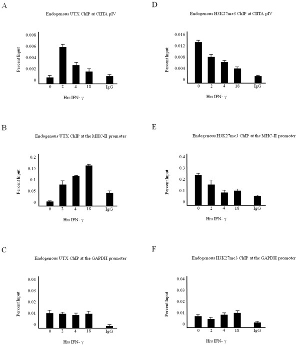
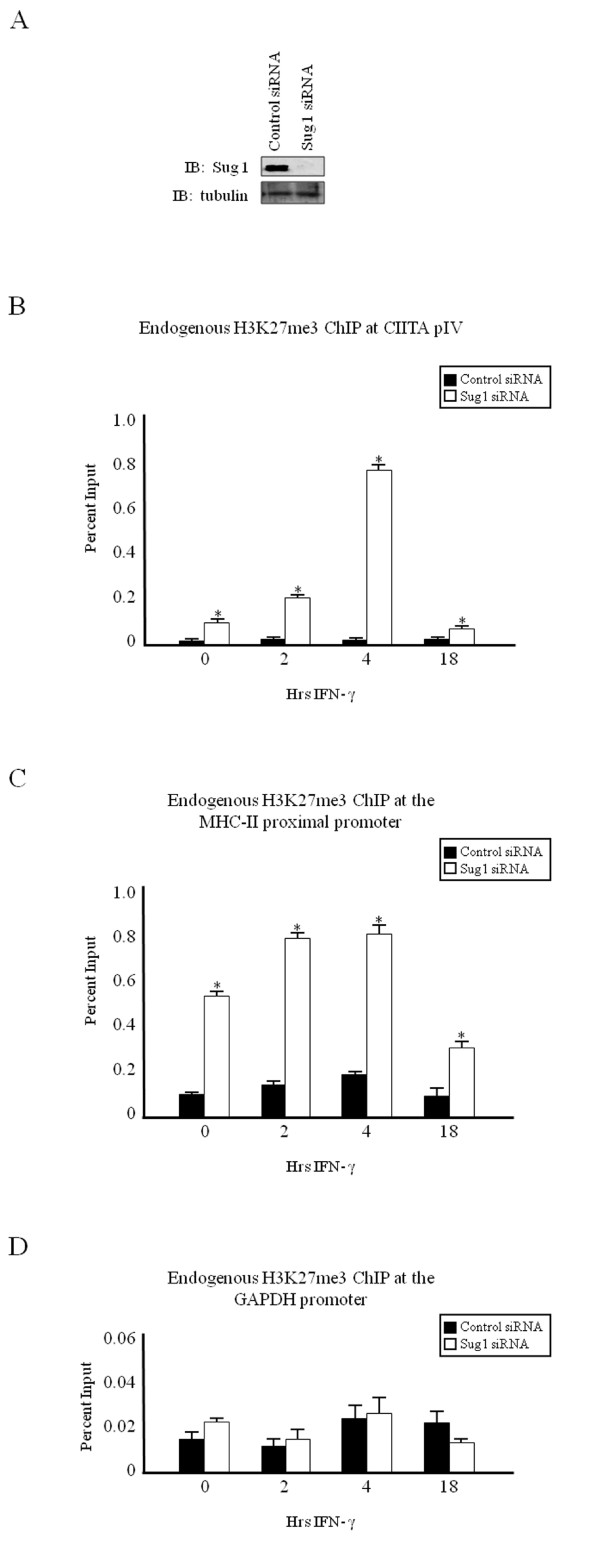
Background: Studies indicate that the 19S proteasome contributes to chromatin reorganization, independent of the role the proteasome plays in protein degradation. We have previously shown that components of the 19S proteasome are crucial for regulating inducible histone activation events in mammalian cells. The 19S ATPase Sug1 binds to histone-remodeling enzymes, and in the absence of Sug1, a subset of activating epigenetic modifications including histone H3 acetylation, H3 lysine 4 trimethylation and H3 arginine 17 dimethylation are inhibited at cytokine-inducible major histocompatibilty complex (MHC)-II and class II transactivator (CIITA) promoters, implicating Sug1 in events required to initiate mammalian transcription.

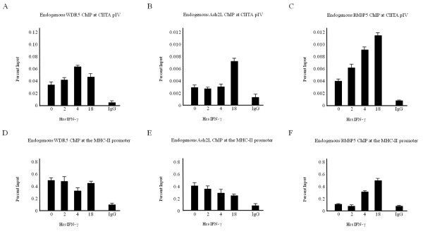
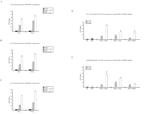
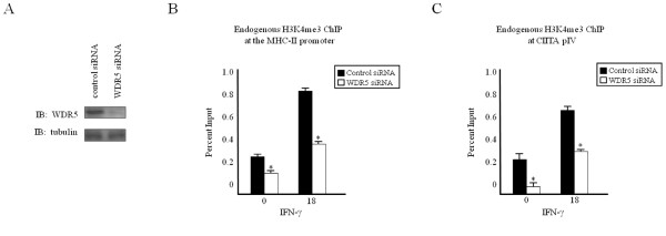
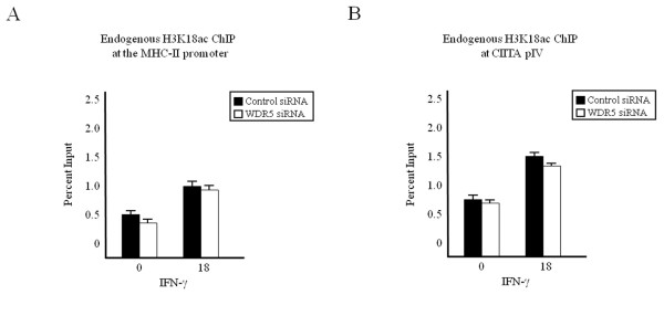
Results: Our previous studies indicate that H3 lysine 4 trimethylation at cytokine-inducible MHC-II and CIITA promoters is dependent on proteolytic-independent functions of 19S ATPases. In this report, we show that multiple common subunits of the mixed lineage leukemia (MLL)/complex of proteins associated with Set I (COMPASS) complexes bind to the inducible MHC-II and CIITA promoters; that overexpressing a single common MLL/COMPASS subunit significantly enhances promoter activity and MHC-II HLA-DRA expression; and that these common subunits are important for H3 lysine 4 trimethylation at MHC-II and CIITA promoters. In addition, we show that H3 lysine 27 trimethylation, which is inversely correlated with H3 lysine 4 trimethylation, is significantly elevated in the presence of diminished 19S ATPase Sug1.

Conclusion: Taken together, these experiments suggest that the 19S proteasome plays a crucial role in the initial reorganization of events enabling the relaxation of the repressive chromatin structure surrounding inducible promoters.


Figures

Figure 1
Figure 1
Common MLL/COMPASS subunits associate with cytokine-inducible genes. (a-c) Several common subunits of MLL/COMPASS associate with CIITA pIV. ChIP assays were carried out in HeLa cells stimulated with IFN-γ for 0 to 18 hours. Lysates were immunoprecipitated with control or endogenous (a) WDR5, (b) Ash2L or (c) RbBP5 antibody. Associated DNA was isolated and analyzed via real-time PCR using primers and probe spanning CIITA pIV. (d-f) Subunits of MLL/COMPASS associate with the MHC-II proximal promoter. ChIP assays were carried out in HeLa cells stimulated with IFN-γ for 0 to 18 hours. Lysates were immunoprecipitated with control or endogenous (d) WDR5, (e) Ash2L or (f) RbBP5 antibody. Associated DNA was isolated and analyzed via real-time PCR using primers and probe spanning the MHC-II proximal promoter. To demonstrate constitutive binding, data are presented as percentage input. IgG isotype control values were 0.001 ± 0.0005 (CIITA pIV) and 0.01 ± 0.007 (MHC-II promoter). Values represent mean ± SEM of (n = 3) independent experiments.
Figure 2
Figure 2
Overexpressing a single common subunit of MLL/COMPASS complexes enhances CIITA transactivity. (A-C) Overexpression of common MLL/COMPASS subunits increases CIITA transactivity at the MHC-II promoter. HeLa cells were transfected with HLA-DRA-Luc, Renilla, pcDNA3, CIITA and (a) WDR5, (b) Ash2L or (c) RbBP5. Luciferase activity is reported as fold activation relative to that of the reporter alone. Luciferase readings were normalized to Renilla activity. Assays were performed in triplicate and values represent mean ± SEM of (n = 3) independent experiments. (d-e) Overexpression of common MLL/COMPASS subunits increases MHC-II transcript levels. HeLa cells were either left untransfected or were transfected with CIITA alone or in combination with WDR5, Ash2L or RbBP5. RNA was prepared using TRIzol reagent and cDNA was generated using gene specific antisense primers for (d) MHC-II HLA-DR and (e) GAPDH. cDNA was quantified via real-time PCR, and data are graphed as fold changes over non-transfected samples. Values represent mean ± SEM of (n = 2) independent experiments. *P < 0.05 vs control siRNA.
Figure 3
Figure 3
Knockdown of a common MLL/COMPASS subunit decreases H3K4me3. (a) WDR5 knockdown efficiently reduces WDR5 expression. HeLa cells were transfected with control or WDR5-specific siRNA, harvested and subjected to western blot analysis for (top) endogenous WDR5 and (bottom) endogenous tubulin. (b-c) WDR5 knockdown decreases H3K4me3. HeLa cells transfected with scrambled control or WDR5-specific siRNA were stimulated with IFN-γ and subjected to ChIP assay. Lysates were immunoprecipitated with control or endogenous H3K4me3 antibody. Associated DNA was isolated and analyzed via real-time PCR as described in Figure 1, using primers and probes specific for (b) the MHC-II promoter or (c) CIITA pIV. Data are presented as percentage input. IgG Isotype control values were 0.06 ± 0.03 (MHC-II promoter) and 0.003 ± 0.002 (CIITA pIV). Values represent mean ± SEM of (n = 3) independent experiments. *P < 0.05 vs control siRNA.
Figure 4
Figure 4
WDR5 knockdown does not affect H3K18ac. (a, b) HeLa cells transfected with scrambled control or WDR5-specific siRNA were stimulated with IFN-γ and subjected to ChIP assay. Lysates were immunoprecipitated with control or endogenous H3K18ac antibody. Associated DNA was isolated and analyzed via real-time PCR as described in Figure 2, using primers and probes specific for (a) MHC-II promoter or (b) CIITA pIV. Data are presented as percentage input. IgG Isotype control values were 0.06 ± 0.03 (MHC-II promoter) and 0.003 ± 0.002 (CIITA pIV). Values represent mean ± SEM of (n = 3) independent experiments.
Figure 5
Figure 5
UTX recruitment correlates with a reduction in H3K27 trimethylation. (a-c) UTX associates with cytokine-inducible promoters. ChIP assays were carried out in HeLa cells stimulated with IFN-γ for 0 to 18 hours. Lysates were immunoprecipitated with control or endogenous UTX antibody. Associated DNA was isolated and analyzed via real-time PCR, using primers and probe spanning (a) CIITA pIV, (b) the MHC-II promoter or (c) the GAPDH promoter. Data are presented as percentage input. Values represent mean ± SEM of (n = 3) independent experiments. (d-f) H3K27 trimethylation was lost upon cytokine stimulation. ChIP assays were carried out in HeLa cells stimulated with IFN-γ for 0 to 18 hours. Lysates were immunoprecipitated with control or endogenous H3K27me3 antibody. Associated DNA was isolated and analyzed via real-time PCR using primers and probe spanning (d) CIITA pIV, (e) the MHC-II promoter or (f) the GAPDH promoter. Data are presented as percentage input. IgG isotype control values were 0.001 ± 0.0002 (CIITA pIV), 0.08 ± 0.02 (MHC-II promoter) and 0.001 ± 0.0009 (GAPDH promoter). Values represent mean ± SEM of (n = 3) independent experiments.
Figure 6
Figure 6
H3K27me3 is enhanced upon Sug1 knockdown. (a) Sug1 siRNA efficiently decreases endogenous Sug1. HeLa cells were transfected with control or Sug1-specific siRNA, harvested and subjected to western blot analysis for (top) endogenous Sug1 and (bottom) endogenous tubulin. (b-d) H3K27me3 is elevated at cytokine-inducible genes upon diminished Sug1 expression. ChIP assays were carried out in HeLa cells stimulated with IFN-γ for 0 to 18 hours. Lysates were immunoprecipitated with control or endogenous H3K27me3 antibody. Associated DNA was isolated and analyzed as in Figure 2 using primers and probe spanning (b) CIITA pIV, (c) the MHC-II proximal promoter and (d) the GAPDH promoter. Data are presented as percentage input. IgG isotype control values were 0.003 ± 0.001 (CIITA pIV), 0.08 ± 0.02 (MHC-II promoter) and 0.01 ± 0.005 (GAPDH promoter). Values represent mean ± SEM of (n = 3) independent experiments.

Similar articles

Cited by

References

    1. Shilatifard A. Chromatin modifications by methylation and ubiquitination: implications in the regulation of gene expression. Annu Rev Biochem. 2006;75:243–269. doi: 10.1146/annurev.biochem.75.103004.142422. - DOI - PubMed
    1. Bernstein BE, Humphrey EL, Erlich RL, Schneider R, Bouman P, Liu JS, Kouzarides T, Schreiber SL. Methylation of histone H3 Lys 4 in coding regions of active genes. Proc Natl Acad Sci USA. 2002;99:8695–8700. doi: 10.1073/pnas.112318199. - DOI - PMC - PubMed
    1. Santos-Rosa H, Schneider R, Bannister AJ, Sherriff J, Bernstein BE, Emre NC, Schreiber SL, Mellor J, Kouzarides T. Active genes are tri-methylated at K4 of histone H3. Nature. 2002;419:407–411. doi: 10.1038/nature01080. - DOI - PubMed
    1. Tsukada Y, Fang J, Erdjument-Bromage H, Warren ME, Borchers CH, Tempst P, Zhang Y. Histone demethylation by a family of JmjC domain-containing proteins. Nature. 2006;439:811–816. doi: 10.1038/nature04433. - DOI - PubMed
    1. Whetstine JR, Nottke A, Lan F, Huarte M, Smolikov S, Chen Z, Spooner E, Li E, Zhang G, Colaiacovo M, Shi Y. Reversal of histone lysine trimethylation by the JMJD2 family of histone demethylases. Cell. 2006;125:467–481. doi: 10.1016/j.cell.2006.03.028. - DOI - PubMed