. 2010 Feb 4:7:9.
doi: 10.1186/1476-9255-7-9.

Histone deacetylase inhibitors induce apoptosis in human eosinophils and neutrophils

Affiliations

Histone deacetylase inhibitors induce apoptosis in human eosinophils and neutrophils

Hannu Kankaanranta et al. J Inflamm (Lond). .

Abstract

Background: Granulocytes are important in the pathogenesis of several inflammatory diseases. Apoptosis is pivotal in the resolution of inflammation. Apoptosis in malignant cells is induced by histone deacetylase (HDAC) inhibitors, whereas HDAC inhibitors do not usually induce apoptosis in non-malignant cells. The aim of the present study was to explore the effects of HDAC inhibitors on apoptosis in human eosinophils and neutrophils.

Methods: Apoptosis was assessed by relative DNA fragmentation assay, annexin-V binding, and morphologic analysis. HDAC activity in nuclear extracts was measured with a nonisotopic assay. HDAC expression was measured by real-time PCR.

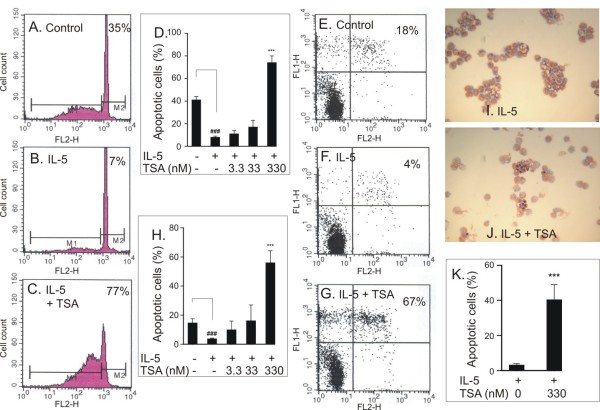
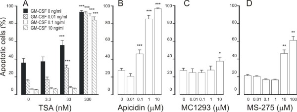
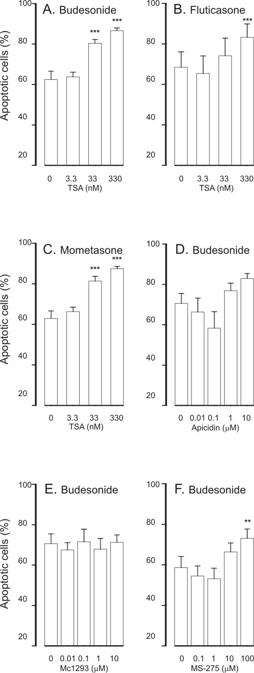
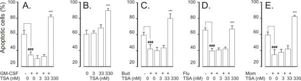
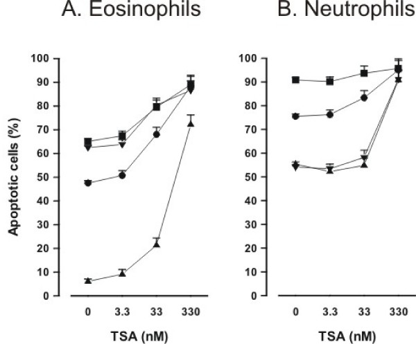
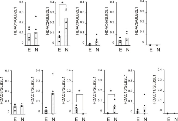
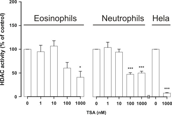
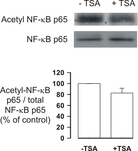
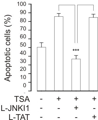
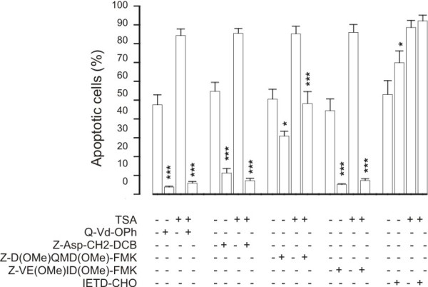
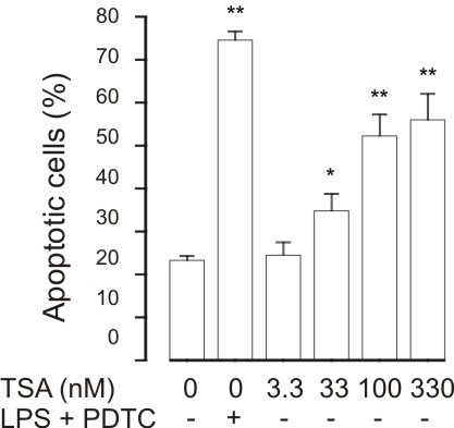
Results: A HDAC inhibitor Trichostatin A (TSA) induced apoptosis in the presence of survival-prolonging cytokines interleukin-5 and granulocyte-macrophage colony stimulating factor (GM-CSF) in eosinophils and neutrophils. TSA enhanced constitutive eosinophil and neutrophil apoptosis. Similar effects were seen with a structurally dissimilar HDAC inhibitor apicidin. TSA showed additive effect on the glucocorticoid-induced eosinophil apoptosis, but antagonized glucocorticoid-induced neutrophil survival. Eosinophils and neutrophils expressed all HDACs at the mRNA level except that HDAC5 and HDAC11 mRNA expression was very low in both cell types, HDAC8 mRNA was very low in neutrophils and HDAC9 mRNA low in eosinophils. TSA reduced eosinophil and neutrophil nuclear HDAC activities by ~50-60%, suggesting a non-histone target. However, TSA did not increase the acetylation of a non-histone target NF-kappaB p65. c-jun-N-terminal kinase and caspases 3 and 6 may be involved in the mechanism of TSA-induced apoptosis, whereas PI3-kinase and caspase 8 are not.

Conclusions: HDAC inhibitors enhance apoptosis in human eosinophils and neutrophils in the absence and presence of survival-prolonging cytokines and glucocorticoids.


Figures

Figure 1
Figure 1
The effect of TSA (330 nM in C, G, J, K) on eosinophil apoptosis in the presence of IL-5 (0.3 ng/ml) as measured by relative DNA fragmentation assay (A-D), Annexin V binding assay (E-H; Annexin V-FITC: FL1-H and propidium iodide: FL2-H)) and morphological analysis (I-K). Figures in top right hand corner represent the percentage of eosinophils showing decreased relative DNA content (A-C) or total percentage of apoptotic eosinophils (all Annexin V-FITC+ve cells) (E-G). In A-C, E-G and I-J a representative of 6 similar experiments is shown. Mean ± SEM, n = 6 (D, H, K). ***P < 0.001 vs. solvent control in the presence of IL-5 and ### P < 0.001 vs. the control in the absence of IL-5 and TSA.
Figure 2
Figure 2
The effect of HDAC inhibitors Trichostatin A (TSA; A), apicidin (B), MC1293 (C) and MS-275 (D) on eosinophil apoptosis in the presence of GM-CSF (in B-D: 0.1 ng/ml). In (A) the black colums indicate the effect of TSA in the absence of GM-CSF. Apoptosis was assessed by flow cytometry measuring the relative DNA fragmentation. *P < 0.05, **P < 0.01 and ***P < 0.001 as compared with the respective control. Mean ± S.E.M., n = 5-6.
Figure 3
Figure 3
The effect of HDAC inhibitors apicidin (A), MC1293 (B) and MS-275 (C) on apoptosis in eosinophils in the absence of survival-prolonging cytokines (ie. spontaneous apoptosis). Apoptosis was assessed by flow cytometry measuring the relative DNA fragmentation in propidium iodide-stained cells. **P < 0.01 and ***P < 0.001 as compared with the respective control in the absence of HDAC inhibitors. Mean ± S.E.M. of 5-6 independent determinations using cells from different donors.
Figure 4
Figure 4
The effect of trichostatin A (A-C) on human eosinophil apoptosis in the presence of budesonide (1 μM; A), fluticasone (1 μM; B) or mometasone (1 μM; C). In (D-F) is shown the effects of HDAC inhibitors apicidin (D), MC1293 (E) and MS-275 (F) on eosinophil apoptosis in the presence of budesonide (1 μM). Apoptosis was assessed by flow cytometry measuring the relative DNA fragmentation in propidium iodide-stained cells. ** indicates P < 0.01 and *** P < 0.001 as compared with the respective control in the absence of HDAC inhibitors. Mean ± S.E.M. of 5-6 independent determinations using cells from different donors. The corresponding percentage of apoptotic cells in the absence of glucocorticoids and HDAC-inhibitors was 49 ± 3 (n = 25).
Figure 5
Figure 5
The effect of Trichostatin A on apoptosis in human neutrophils in the presence (A) or absence (B) of the survival-prolonging cytokine GM-CSF (10 ng/ml). In (C-E) is shown the effect of trichostatin A on human neutrophil apoptosis in the presence of budesonide (1 μM; C), fluticasone (1 μM; D) or mometasone (1 μM; E). Apoptosis was assessed by flow cytometry measuring the relative DNA fragmentation assay. *** P < 0.001 as compared with the respective control in the absence of HDAC inhibitors. ### P < 0.001 as compared with the respective control in the absence of HDAC inhibitors and GM-CSF or glucocorticoids. Mean ± S.E.M. of 6 independent determinations using cells from different donors.
Figure 6
Figure 6
Concentration-response curves of TSA in eosinophils (A) and neutrophils (B) in the absence (black circle) and presence of survival-prolonging cytokines (black up-pointing triangle; IL-5 0.3 ng/ml in eosinophils and GM-CSF 10 ng/ml in neutrophils), budesonide (black down-pointing triangle; 1 μM) or Fas (black square; 100 ng/ml). Apoptosis was assessed by flow cytometry measuring the relative DNA fragmentation (A) or Annexin V-binding (B). Eosinophils or neutrophils were isolated and concentration-response curves in the absence or presence of cytokines, budesonide or Fas were prepared simultaneously from the cells of the same donor. Mean ± S.E.M. of 6 independent determinations using cells from different donors.
Figure 7
Figure 7
The expression of histone deacetylases (HDAC) 1-11 in human eosinophils (black circle; E) and neutrophils (black up-pointing triangle; N). HDAC mRNA levels were normalized against GLB2L1 mRNA. Total mRNA from eosinophils (n = 4) and neutrophils (n = 5) was extracted and subjected to RT-PCR. *P < 0.05 for the difference between eosinophils and neutrophils.
Figure 8
Figure 8
The effect of HDAC inhibitor Trichostatin A (TSA) on HDAC activity in nuclear extracts isolated from human eosinophils (n = 6) and neutrophils (n = 5). For comparison is shown the effect of TSA on HDAC activity in HeLa nuclear extracts (n = 6). Nuclear extracts were prepared and HDAC activity was measured as described in materials and methods. HDAC activity in the absence of TSA was set as 100%. *P < 0.05 and *** P < 0.001 as compared with the respective control in the absence of TSA. Mean ± S.E.M.
Figure 9
Figure 9
The effect of TSA (330 nM) on the expression of acetyl-NF-kB p65 (Lys310). Human eosinophils were treated with solvent or TSA for 1 h and immunoblots were run using antibodies against acetyl-NF-kB p65 and total NF-kB p65. The chemiluminescent signal was quantified as described under Materials and Methods. Acetyl-NF-kB p65 values were normalized to NF-kB p65 values and the value in the absence of TSA was set as 100%. Results are expressed as mean ± S.E.M., n = 5.
Figure 10
Figure 10
The effect of the c-jun-N-terminal kinase inhibitor L-JNKI1 (10 μM) on TSA (330 nM)-induced human eosinophil apoptosis. Apoptosis was assessed by the relative DNA fragmentation assay. Each data point represents the mean ± SEM of 6 independent determinations using eosinophils from different donors. *** indicates p < 0.001 as compared with the respective control (10 μM L-TAT or solvent).
Figure 11
Figure 11
The effect of caspase inhibitors on TSA (330 nM)-induced human eosinophil apoptosis. The concentrations used were: Q-Vd-Oph (20 μM), Z-Asp-CH2-DCB and IETD-CHO (100 μM) and Z-D(OMe)QMD(OMe)-FMK and Z-VE(OMe)ID(OMe)-FMK (200 μM). Apoptosis was assessed by the relative DNA fragmentation assay. Each data point represents the mean ± SEM of 6-7 independent determinations using eosinophils from different donors. * indicates p < 0.05 and *** p < 0.001 as compared with the respective control in the absence of caspase inhibitors.
Figure 12
Figure 12
The effect of HDAC inhibitor Trichostatin A (TSA) on apoptosis in J774.2 macrophages during culture for 24 h. For comparison, is shown the effect of a known inducer of apoptosis in J774 macrophages, i.e. combination of LPS (10 ng/ml) and PDTC (100 μM). Apoptosis was assessed by flow cytometry measuring the percentage of Annexin V-positive cells. *P < 0.05 and **P < 0.01 as compared with the respective control. Mean ± S.E.M., n = 12.

Similar articles

Cited by

References

    1. Watt AP, Schock BC, Ennis M. Neutrophils and eosinophils: clinical implications of their appearance, presence and disappearance in asthma and COPD. Curr Drug Targets-Inflammation Allergy. 2005;4:415–423. doi: 10.2174/1568010054526313. - DOI - PubMed
    1. Walker A, Ward C, Taylor EL, Dransfield I, Hart SP, Haslett C, Rossi AG. Regulation of neutrophil apoptosis and removal of apoptotic cells. Curr Drug Targets-Inflammation Allergy. 2005;4:447–454. doi: 10.2174/1568010054526278. - DOI - PubMed
    1. Kankaanranta H, Moilanen E, Zhang X. Pharmacological regulation of human eosinophil apoptosis. Curr Drug Targets-Inflammation Allergy. 2005;4:433–445. doi: 10.2174/1568010054526395. - DOI - PubMed
    1. Wedi B, Raap U, Lewrick H, Kapp A. Delayed eosinophil programmed cell death in vitro: a common feature of inhalant allergy and extrinsic and intrinsic atopic dermatitis. J Allergy Clin Immunol. 1997;100:536–43. doi: 10.1016/S0091-6749(97)70147-7. - DOI - PubMed
    1. Kankaanranta H, Lindsay MA, Giembycz MA, Zhang X, Moilanen E, Barnes PJ. Delayed eosinophil apoptosis in asthma. J Allergy Clin Immunol. 2000;106:77–83. doi: 10.1067/mai.2000.107038. - DOI - PubMed