. 2010 Feb 5;8(1):1.
doi: 10.1186/1479-0556-8-1.

Optimised electroporation mediated DNA vaccination for treatment of prostate cancer

Affiliations

Optimised electroporation mediated DNA vaccination for treatment of prostate cancer

Sarfraz Ahmad et al. Genet Vaccines Ther. .

Abstract

Background: Immunological therapies enhance the ability of the immune system to recognise and destroy cancer cells via selective killing mechanisms. DNA vaccines have potential to activate the immune system against specific antigens, with accompanying potent immunological adjuvant effects from unmethylated CpG motifs as on prokaryotic DNA. We investigated an electroporation driven plasmid DNA vaccination strategy in animal models for treatment of prostate cancer.

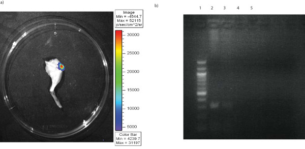
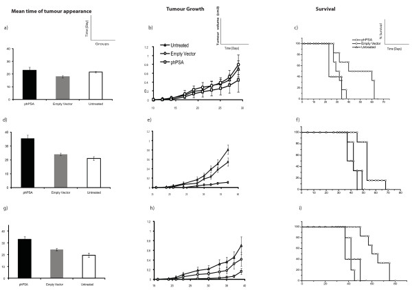
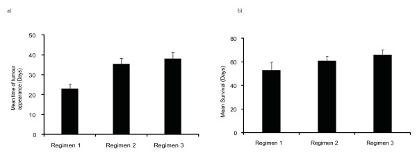
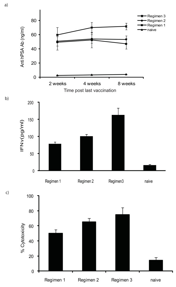
Methods: Plasmid expressing human PSA gene (phPSA) was delivered in vivo by intra-muscular electroporation, to induce effective anti-tumour immune responses against prostate antigen expressing tumours. Groups of male C57 BL/6 mice received intra-muscular injections of phPSA plasmid. For phPSA delivery, quadriceps muscle was injected with 50 microg plasmid. After 80 seconds, square-wave pulses were administered in sequence using a custom designed pulse generator and a custom-designed applicator with 2 needles placed through the skin central to the muscle. To determine an optimum treatment regimen, three different vaccination schedules were investigated. In a separate experiment, the immune potential of the phPSA vaccine was further enhanced with co- administration of synthetic CpG rich oligonucleotides. One week after last vaccination, the mice were challenged subcutaneously with TRAMPC1/hPSA (prostate cancer cell line stably expressing human PSA) and tumour growth was monitored. Serum from animals was examined by ELISA for anti-hPSA antibodies and for IFN gamma. Histological assessment of the tumours was also carried out. In vivo and in vitro cytotoxicity assays were performed with splenocytes from treated mice.

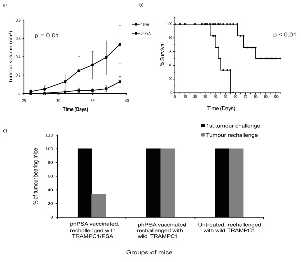
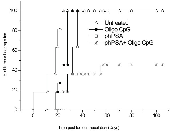
Results: The phPSA vaccine therapy significantly delayed the appearance of tumours and resulted in prolonged survival of the animals. Four-dose vaccination regimen provided optimal immunological effects. Co - administration of the synthetic CpG with phPSA increased anti-tumour responses, preventing tumour occurrence in 54% of treated animals. Vaccination with phPSA resulted in anti-hPSA Abs production and a significant production of IFN gamma was observed in immunised animals (p < 0.05). Immune responses were tumour specific and were transferable in adoptive T cell transfer experiments.

Conclusions: This phPSA plasmid electroporation vaccination strategy can effectively activate tumour specific immune responses. Optimisation of the approach indicated that a four-dose regimen provided highest tumour protection. In vivo electroporation mediated vaccination is a safe and effective modality for the treatment of prostate cancer and has a potential to be used as a neo-adjuvant or adjuvant therapy.


Figures

Figure 1
Figure 1
Schematic representation of vaccination schedules. Regimen1 involved two vaccinations (day 0 and 14) with subsequent tumour challenge on day 15. Regimen 2 involved vaccinations on day 0, 7, and tumour challenge on day 14. In regimen 3, four doses of vaccine were given on day 0, 7, 14, and 21 followed by tumour challenge on day 28.
Figure 2
Figure 2
Electroporation mediate plasmid transfection of quadriceps. a) In vivo muscle transfection by EP was assessed by luciferase activity in resected leg 72 h post transfection (imaged for 1 min using an intensified CCD camera (IVIS Imaging System, Xenogen). b) RT-PCR analysis of mRNA expression of hPSA in muscle. hPSA was only detected in muscles electroporated with phPSA (lane 1 100 bp marker, lane 2 phPSA transfected muscle (sample a), lane 3 phPSA transfected muscle (sample b), lane 4 empty vector transfected muscle, lane 5 untreated muscle).
Figure 3
Figure 3
Tumour protective effects of the various vaccination regimens. (n = 6) Regimen 1 - a) Time of tumour appearance - the mean time of tumour appearance was comparable in various groups (p = 0.07). b) Representative tumour growth curve - phPSA immunised mice had low tumour volumes but the difference was not significant (p vs empty vector = 0.34, vs untreated = 0.27). c) Representative Kaplan Meyer survival curve - mean survival in the immunised group was significantly prolonged (p vs empty vector = 0.04, vs untreated = 0.03). Regimen 2 - d) Time of tumour appearance - the phPSA treated mice remained tumours free for prolonged period of time (p < 0.01). e) Representative tumour growth curve - tumour growth was retarded in the immunised group. The tumour volumes were significantly lower than the untreated group at all time points (p = 0.04), but not when compared with empty vector group (p = 0.07). f) Representative Kaplan Meyer survival curve - immunisation with phPSA provided significant prolonged survival of the treated mice (p vs empty vector = 0.01, vs untreated = 0.01). Regimen 3 - g) Time of tumour appearance - mean time of tumour appearance was delayed significantly as compared to both control groups (p < 0.01). h) Representative tumour growth curve - tumour growth was significantly retarded in phPSA immunised group (p vs empty vector = 0.04, vs untreated = 0.01). i) Representative Kaplan Meyer survival curve - average survival in immunise group was significantly prolonged (p vs empty vector < 0.01, vs untreated < 0.01). Data are expressed as means ± SEM.
Figure 4
Figure 4
Comparison of vaccination regimens. a) Time of tumour appearance in various vaccinated groups. The regimen 2 and 3 resulted in prolonged tumour free periods (p regimen 1 vs 2 < 0.01, regimen 1 vs 3 < 0.01, and regimen 2 vs 3 = 0.4). b) Mean survival in various vaccination regimens (p regimen 1 vs 2 = 0.4, regimen1 vs 3 = 0.04, regimen 2 vs 3 = 0.04). Data shown only for the phPSA vaccinated mice in all three regimens. Errors bars represent SE.
Figure 5
Figure 5
Induction of protective anti-tumour immunity. a) Levels of anti-hPSA antibodies at various time points from both vaccinated and from naive mice. Elevated levels of the anti hPSA antibodies were found in the serum from the immunised mice. Regimen 3 resulted in higher levels at all time points. Errors bars represent SE. b) IFNγ production - at study end point in various groups (n = 6), supernatants from the stimulated (48 hours) splenocytes collected and tested for the production of IFNγ. Higher levels of IFNγ were detected in all groups as compared to naive, while regimen 3 resulted in a much higher IFNγ production (p vs regimen 1 < 0.01, vs regimen 2 = 0.01). Regimen 2 levels were also significantly higher as compared to regimen 1 (p = 0.03). c) In vitro augmentation of the cytolytic activities of the splenocytes after immunisation - specific cytotoxicity was greatest at an effector target ratio of 20:1 in all vaccination schedules. Maximum cytotoxicity was observed in regimen 3. The % cytotoxicity was significantly higher than naive for all regimens, but differences between regimens were not statistically different (p > 0.05) (p vs regimen 3 = 0.02, vs regimen 2 = 0.01, vs regimen 1 = 0.03). Errors bars represent SE.
Figure 6
Figure 6
In vivo adoptive transfer of lymphocytes and antigen specific response. a) Low tumour volumes were observed in mice (n = 6) receiving splenocytes from immunised group and this slow growth of the tumours provided a prolonged survival. b). Data are expressed as means ± SEM. c) Antigen specific responses - groups of mice (n = 6) were immunised (regimen 3) and challenged s.c. (1st tumour challenge) either with wild TRAMPC1 or transfected TRAMPC1 (TRAMPC1/hPSA). After surgical excision of the tumours and 30 days period of observation, mice were rechallenged (Tumour rechallenge), either with wild type TRAMPC1 or TRAMPC1/hPSA cells. The vaccine response was antigen specific, as on re-challenge tumour protection was only observed in mice with neo-adjuvant immunisation and rechallenge with TRAMPC1/hPSA. Only 33% mice developed tumour after rechallenge, while remaining mice remained tumour free for more than 100 days post rechallenge with TRAMPC1/hPSA.
Figure 7
Figure 7
Time of tumour development. Co-administration of the synthetic oligo CpG with phPSA (regimen 3) resulted in delayed onset of tumours (n = 11). 54% mice (6 out of 11) remained tumour free for > 100 days post s.c. tumour inoculation. The tumour development in remaining five mice was delayed as compared to other groups.

References

    1. Jemal A, Siegel R, Ward E, Murray T, Xu J, Thun MJ. Cancer statistics, 2007. CA Cancer J Clin. 2007;57:43–66. doi: 10.3322/canjclin.57.1.43. - DOI - PubMed
    1. Fournier P, Schirrmacher V. Randomized clinical studies of anti-tumor vaccination: state of the art in 2008. Expert Rev Vaccines. 2009;8:51–66. doi: 10.1586/14760584.8.1.51. - DOI - PubMed
    1. Blades RA, Keating PJ, McWilliam LJ, George NJ, Stern PL. Loss of HLA class I expression in prostate cancer: implications for immunotherapy. Urology. 1995;46:681–686. doi: 10.1016/S0090-4295(99)80301-X. - DOI - PubMed
    1. Mukherji B, Chakraborty NG. Immunobiology and immunotherapy of melanoma. Curr Opin Oncol. 1995;7:175–184. doi: 10.1097/00001622-199503000-00014. - DOI - PubMed
    1. Zier K, Gansbacher B, Salvadori S. Preventing abnormalities in signal transduction of T cells in cancer: the promise of cytokine gene therapy. Immunol Today. 1996;17:39–45. doi: 10.1016/0167-5699(96)80567-6. - DOI - PubMed