

Capillary hemangioma as a rare benign tumor of the oral cavity: a case report
- PMID: 20181211
- PMCID: PMC2827094
- DOI: 10.1186/1757-1626-0002-0000008622
Capillary hemangioma as a rare benign tumor of the oral cavity: a case report
Abstract
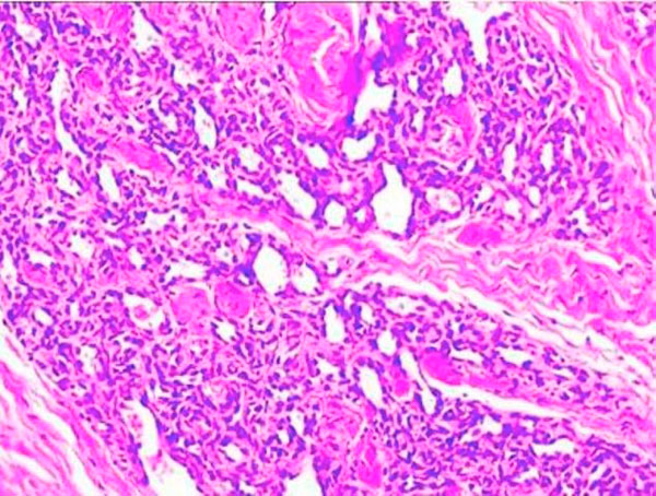
Introduction: Hemangioma is a relatively common benign proliferation of blood vessels that primarily develops during childhood. Two main forms of hemangioma recognized: capillary and cavernous. The capillary form presents as a flat area consisting of numerous small capillaries. Cavernous hemangioma appears as an elevated lesion of a deep red color, and consists of large dilated sinuses filled with blood. The purpose of the study was to report the case of a capillary hemangioma in a patient and to describe the successful treatment of this case.
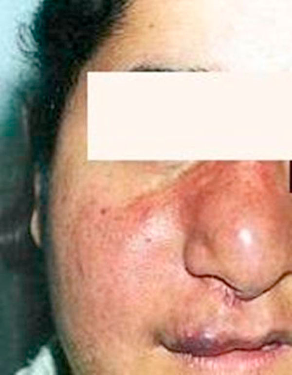
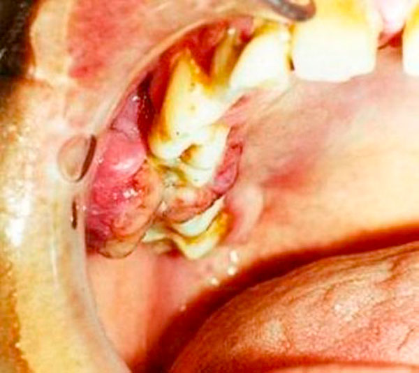
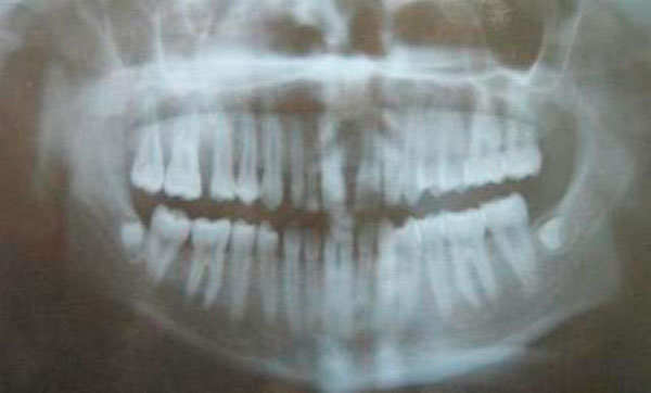
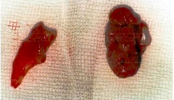
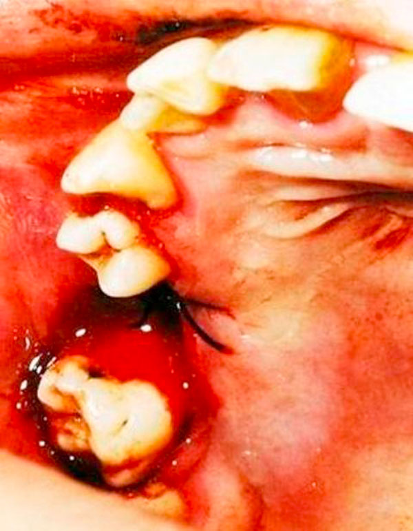
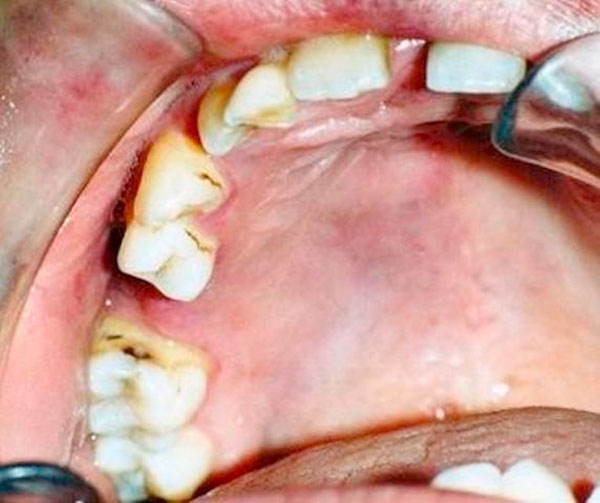
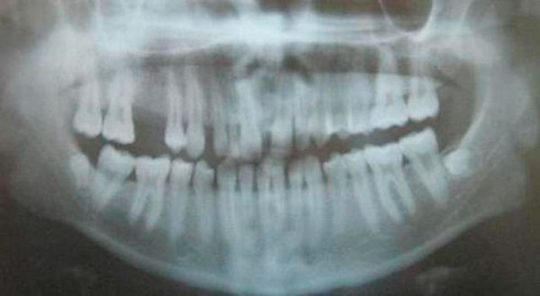
Case presentation: The patient was a 19-year-old female who presented herself to the Atatürk University, Faculty of Dentistry, Department of Periodontology, with the complaint of bleeding and slowly enlarging mass on the upper right molar region. The lesion was diagnosed as capillary hemangioma after clinical examination and biopsy. Treatment consisted of scaling, root planning and surgical excision. Four months after surgery healing was occurred and two years later area of the lesion appeared completely normal as clinically.
Conclusions: The surface is highly keratinized and no further growth was evidenced during the two year of follow-up. Early detection and biopsy is necessary to determine the clinical behavior of the tumor and potential dentoalveolar complications.
Figures
Similar articles
-
A cavernous hemangioma of the tongue base presenting as an ectopic thyroid: A case report.Ann Med Surg (Lond). 2020 Oct 23;60:115-120. doi: 10.1016/j.amsu.2020.10.053. eCollection 2020 Dec. Ann Med Surg (Lond). 2020. PMID: 33145019 Free PMC article.
-
Intranodal capillary-cavernous hemangioma: Report of a very rare case.SAGE Open Med Case Rep. 2019 May 2;7:2050313X19846710. doi: 10.1177/2050313X19846710. eCollection 2019. SAGE Open Med Case Rep. 2019. PMID: 31105947 Free PMC article.
-
Characteristics of a pediatric patient with a capillary hemangioma of the palatal mucosa: a case report.Pediatr Dent. 2007 May-Jun;29(3):239-42. Pediatr Dent. 2007. PMID: 17688022
-
Lymphocyte-rich capillary-cavernous hemangioma of the mitral valve: a case report and review of the literature.Cardiovasc Pathol. 2017 May-Jun;28:59-63. doi: 10.1016/j.carpath.2017.03.003. Epub 2017 Mar 10. Cardiovasc Pathol. 2017. PMID: 28334596 Review.
-
[Neoplastic space-occupying lesions of the orbit. I. Review; hemangioma, lymphangioma and embryonal rhabdomyosarcoma].Klin Monbl Augenheilkd. 1992 Nov;201(5):291-301. doi: 10.1055/s-2008-1045906. Klin Monbl Augenheilkd. 1992. PMID: 1479785 Review. German.
Cited by
-
Defocused irradiation mode of diode laser for conservative treatment of oral hemangioma.J Lasers Med Sci. 2013 Summer;4(3):147-50. J Lasers Med Sci. 2013. PMID: 25606323 Free PMC article.
-
Transoral surgery with Thunderbeat© for hemangioma of the tongue base: A novel procedure. Case report.Int J Surg Case Rep. 2024 Mar;116:109385. doi: 10.1016/j.ijscr.2024.109385. Epub 2024 Feb 10. Int J Surg Case Rep. 2024. PMID: 38350379 Free PMC article.
-
A giant compound hemangioma of lower lip.J Oral Maxillofac Pathol. 2012 Sep;16(3):438-40. doi: 10.4103/0973-029X.102512. J Oral Maxillofac Pathol. 2012. PMID: 23248483 Free PMC article.
-
Lobular Capillary Hemangioma of the Buccal Mucosa: A Rare Presentation.Cureus. 2024 Jul 31;16(7):e65904. doi: 10.7759/cureus.65904. eCollection 2024 Jul. Cureus. 2024. PMID: 39219971 Free PMC article.
-
A cavernous hemangioma of the tongue base presenting as an ectopic thyroid: A case report.Ann Med Surg (Lond). 2020 Oct 23;60:115-120. doi: 10.1016/j.amsu.2020.10.053. eCollection 2020 Dec. Ann Med Surg (Lond). 2020. PMID: 33145019 Free PMC article.
References
-
- Enzinger FM, Weiss SW. Soft tissue tumors. 3. Mosby; 1995. pp. 581–586.
-
- Silverman RA. Hemangiomas and vascular malformations. Pediatr Clin North Am. 1991;38:811–834. - PubMed
LinkOut - more resources
Full Text Sources
