

Premature infant swallowing: patterns of tongue-soft palate coordination based upon videofluoroscopy
- PMID: 20181397
- PMCID: PMC2844905
- DOI: 10.1016/j.infbeh.2009.10.001
Premature infant swallowing: patterns of tongue-soft palate coordination based upon videofluoroscopy
Abstract
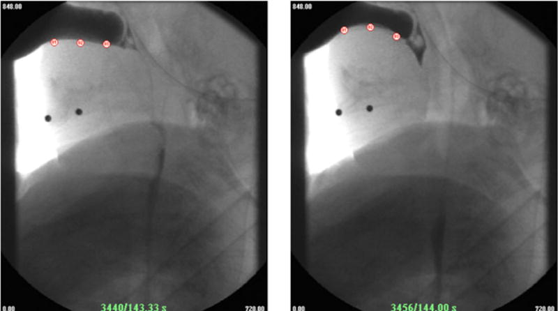
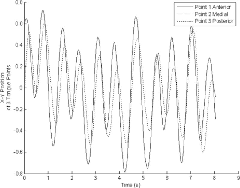
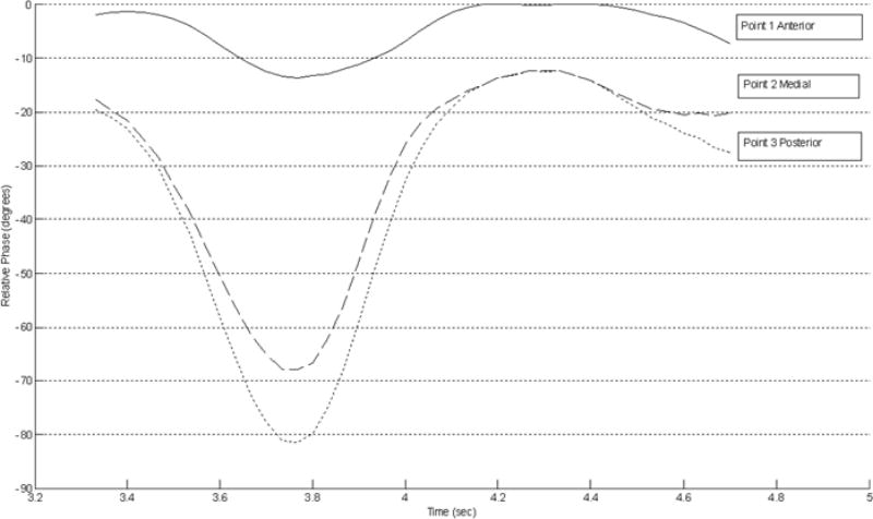
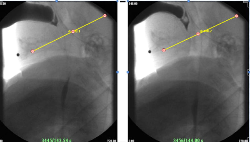
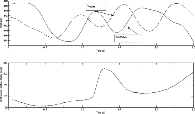
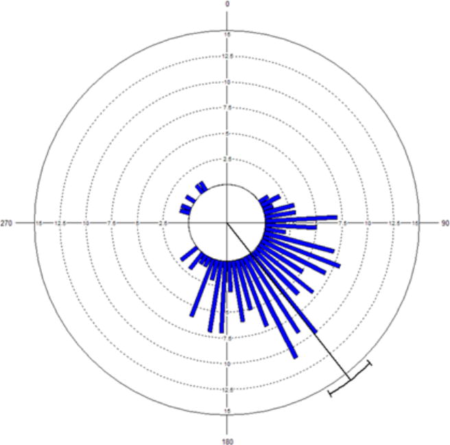
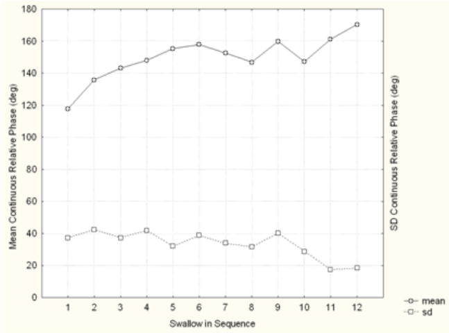
Coordination between movements of individual tongue points, and between soft palate elevation and tongue movements, were examined in 12 prematurely born infants referred from hospital NICUs for videofluoroscopic swallow study (VFSS) due to poor oral feeding and suspicion of aspiration. Detailed post-evaluation kinematic analysis was conducted by digitizing images of a lateral view of digitally superimposed points on the tongue and soft palate. The primary measure of coordination was continuous relative phase of the time series created by movements of points on the tongue and soft palate over successive frames. Three points on the tongue (anterior, medial, and posterior) were organized around a stable in-phase pattern, with a phase lag that implied an anterior to posterior direction of motion. Coordination between a tongue point and a point on the soft palate during lowering and elevation was close to anti-phase at initiation of the pharyngeal swallow. These findings suggest that anti-phase coordination between tongue and soft palate may reflect the process by which the tongue is timed to pump liquid by moving it into an enclosed space, compressing it, and allowing it to leave by a specific route through the pharynx.
Copyright 2009 Elsevier Inc. All rights reserved.
Figures
Similar articles
-
Preterm infant swallowing of thin and nectar-thick liquids: changes in lingual-palatal coordination and relation to bolus transit.Dysphagia. 2013 Jun;28(2):234-44. doi: 10.1007/s00455-012-9440-y. Epub 2013 Jan 1. Dysphagia. 2013. PMID: 23274694 Free PMC article.
-
Tongue pressure patterns during water swallowing.Dysphagia. 2010 Mar;25(1):11-9. doi: 10.1007/s00455-009-9223-2. Epub 2009 Jun 30. Dysphagia. 2010. PMID: 19568810
-
Effects of respiration on soft palate movement in feeding.J Dent Res. 2010 Dec;89(12):1401-6. doi: 10.1177/0022034510377336. Epub 2010 Sep 1. J Dent Res. 2010. PMID: 20811071 Free PMC article.
-
Quantitative description of eustachian tube movements during swallowing as visualized by transnasal videoendoscopy.JAMA Otolaryngol Head Neck Surg. 2015 Feb;141(2):160-8. doi: 10.1001/jamaoto.2014.3002. JAMA Otolaryngol Head Neck Surg. 2015. PMID: 25474183
-
Properties of tissues surrounding the upper airway.Sleep. 1996 Dec;19(10 Suppl):S170-4. doi: 10.1093/sleep/19.suppl_10.170. Sleep. 1996. PMID: 9085502 Review.
Cited by
-
Variability in Swallowing Biomechanics in Infants with Feeding Difficulties: A Videofluoroscopic Analysis.Dysphagia. 2022 Dec;37(6):1740-1747. doi: 10.1007/s00455-022-10436-2. Epub 2022 Mar 17. Dysphagia. 2022. PMID: 35298686 Free PMC article.
-
Verification of Reliability and Validity of the Feeding and Swallowing Scale for Premature Infants (FSSPI).Ann Rehabil Med. 2017 Aug;41(4):631-637. doi: 10.5535/arm.2017.41.4.631. Epub 2017 Aug 31. Ann Rehabil Med. 2017. PMID: 28971048 Free PMC article.
-
Reduced Coordination of Hyolaryngeal Elevation and Bolus Movement in a Pig Model of Preterm Infant Swallowing.Dysphagia. 2020 Apr;35(2):334-342. doi: 10.1007/s00455-019-10033-w. Epub 2019 Jul 11. Dysphagia. 2020. PMID: 31297599 Free PMC article.
-
Swallowing Analyses of Neonates and Infants in Breastfeeding and Bottle-feeding: Impact on Videofluoroscopy Swallow Studies.Int Arch Otorhinolaryngol. 2019 Jul;23(3):e343-e353. doi: 10.1055/s-0039-1677753. Epub 2019 May 28. Int Arch Otorhinolaryngol. 2019. PMID: 31360257 Free PMC article.
-
Preterm infant swallowing of thin and nectar-thick liquids: changes in lingual-palatal coordination and relation to bolus transit.Dysphagia. 2013 Jun;28(2):234-44. doi: 10.1007/s00455-012-9440-y. Epub 2013 Jan 1. Dysphagia. 2013. PMID: 23274694 Free PMC article.
References
-
- Bosma JF, Hepburn LG, Josell SD, Baker K. Ultrasound demonstration of tongue motions during suckle feeding. Developmental Medicine and Child Neurology. 1990;32:223–229. - PubMed
-
- Bu’Lock F, Woolridge MW, Baum JD. Development of coordination of suckling, swallowing, and breathing: Ultrasound study of term and preterm infants. Develop Med Child Neurol. 1990;32:669–678. - PubMed
-
- Cohen AH. Effects of oscillator frequency on phase-locking in the lamprey central pattern generator. Journal of Neuroscience Methods. 1987;21:113–125. - PubMed
-
- Dodds WJ. The physiology of swallowing. Dysphagia. 1989;3:171–178. - PubMed
MeSH terms
Grants and funding
LinkOut - more resources
Full Text Sources
Other Literature Sources
Medical
