

Interaction between Ephrins and mGlu5 metabotropic glutamate receptors in the induction of long-term synaptic depression in the hippocampus
- PMID: 20181581
- PMCID: PMC6633947
- DOI: 10.1523/JNEUROSCI.4834-09.2010
Interaction between Ephrins and mGlu5 metabotropic glutamate receptors in the induction of long-term synaptic depression in the hippocampus
Abstract
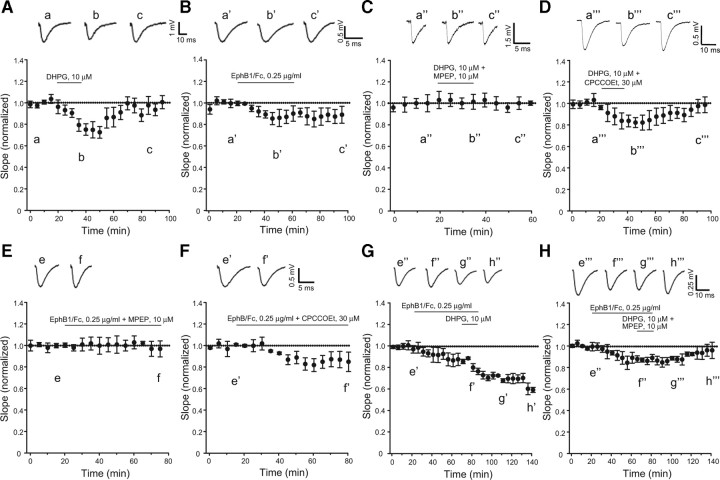
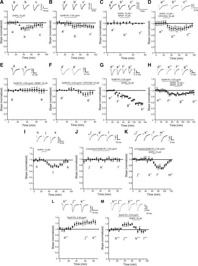
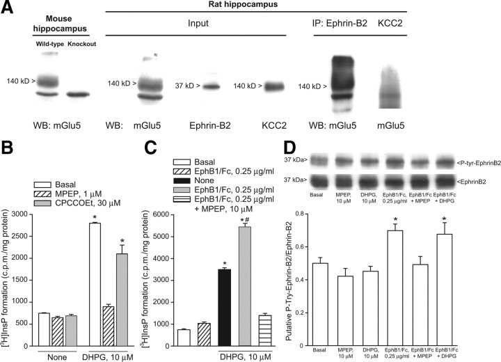
We applied the group-I metabotropic glutamate (mGlu) receptor agonist, 3,5-dihydroxyphenylglycine (DHPG), to neonatal or adult rat hippocampal slices at concentrations (10 microM) that induced a short-term depression (STD) of excitatory synaptic transmission at the Schaffer collateral/CA1 synapses. DHPG-induced STD was entirely mediated by the activation of mGlu5 receptors because it was abrogated by the mGlu5 receptor antagonist, MPEP [2-methyl-6-(phenylethynyl)pyridine], but not by the mGlu1 receptor antagonist, CPCCOEt [7-(hydroxyimino)cyclopropa[b]chromen-1a-carboxylate ethyl ester]. Knowing that ephrin-Bs functionally interact with group-I mGlu receptors (Calò et al., 2005), we examined whether pharmacological activation of ephrin-Bs could affect DHPG-induced STD. We activated ephrin-Bs using their cognate receptor, EphB1, under the form of a preclustered EphB1/Fc chimera. Addition of clustered EphB1/Fc alone to the slices induced a small but nondecremental depression of excitatory synaptic transmission, which differed from the depression induced by 10 microM DHPG. Surprisingly, EphB1/Fc-induced synaptic depression was abolished by MPEP (but not by CPCCOEt) suggesting that it required the endogenous activation of mGlu5 receptors. In addition, coapplication of DHPG and EphB1/Fc, resulted in a large and nondecremental long-term depression. The effect of clustered EphB1/Fc was specific because it was not mimicked by unclustered EphB1/Fc or clustered EphA1/Fc. These findings raise the intriguing possibility that changes in synaptic efficacy mediated by mGlu5 receptors are under the control of the ephrin/Eph receptor system, and that the neuronal actions of ephrins can be targeted by drugs that attenuate mGlu5 receptor signaling.
Figures

Similar articles
-
Activation of metabotropic glutamate 5 (mGlu5) receptors induces spontaneous excitatory synaptic currents in layer V pyramidal cells of the rat prefrontal cortex.Neurosci Lett. 2008 Sep 19;442(3):239-43. doi: 10.1016/j.neulet.2008.06.083. Epub 2008 Jul 4. Neurosci Lett. 2008. PMID: 18621097 Free PMC article.
-
Molecular mechanisms of group I metabotropic glutamate receptor mediated LTP and LTD in basolateral amygdala in vitro.Psychopharmacology (Berl). 2017 Feb;234(4):681-694. doi: 10.1007/s00213-016-4503-7. Epub 2016 Dec 28. Psychopharmacology (Berl). 2017. PMID: 28028604
-
Metabotropic glutamate receptor 1 activity generates persistent, N-methyl-D-aspartate receptor-dependent depression of hippocampal pyramidal cell excitability.Eur J Neurosci. 2009 Jun;29(12):2347-62. doi: 10.1111/j.1460-9568.2009.06780.x. Epub 2009 May 29. Eur J Neurosci. 2009. PMID: 19490024
-
Group I mGluR Induced LTD of NMDAR-synaptic Transmission at the Schaffer Collateral but not Temperoammonic Input to CA1.Curr Neuropharmacol. 2016;14(5):435-40. doi: 10.2174/1570159x13666150615221502. Curr Neuropharmacol. 2016. PMID: 27296639 Free PMC article. Review.
-
Dysregulation of group-I metabotropic glutamate (mGlu) receptor mediated signalling in disorders associated with Intellectual Disability and Autism.Neurosci Biobehav Rev. 2014 Oct;46 Pt 2(Pt 2):228-41. doi: 10.1016/j.neubiorev.2014.02.003. Epub 2014 Feb 15. Neurosci Biobehav Rev. 2014. PMID: 24548786 Free PMC article. Review.
Cited by
-
Altered genome-wide hippocampal gene expression profiles following early life lead exposure and their potential for reversal by environmental enrichment.Sci Rep. 2022 Jul 25;12(1):11937. doi: 10.1038/s41598-022-15861-9. Sci Rep. 2022. PMID: 35879375 Free PMC article.
-
Chronic corticosterone administration down-regulates metabotropic glutamate receptor 5 protein expression in the rat hippocampus.Neuroscience. 2010 Sep 15;169(4):1567-74. doi: 10.1016/j.neuroscience.2010.06.023. Epub 2010 Jun 23. Neuroscience. 2010. PMID: 20600666 Free PMC article.
-
Transduction of group I mGluR-mediated synaptic plasticity by β-arrestin2 signalling.Nat Commun. 2016 Nov 25;7:13571. doi: 10.1038/ncomms13571. Nat Commun. 2016. PMID: 27886171 Free PMC article.
-
A novel form of low-frequency hippocampal mossy fiber plasticity induced by bimodal mGlu1 receptor signaling.J Neurosci. 2011 Nov 23;31(47):16897-906. doi: 10.1523/JNEUROSCI.1264-11.2011. J Neurosci. 2011. PMID: 22114260 Free PMC article.
-
NMDA receptors, mGluR5, and endocannabinoids are involved in a cascade leading to hippocampal long-term depression.Neuropsychopharmacology. 2012 Feb;37(3):609-17. doi: 10.1038/npp.2011.243. Epub 2011 Oct 12. Neuropsychopharmacology. 2012. PMID: 21993209 Free PMC article.
References
-
- Ango F, Prézeau L, Muller T, Tu JC, Xiao B, Worley PF, Pin JP, Bockaert J, Fagni L. Agonist-independent activation of metabotropic glutamate receptors by the intracellular protein Homer. Nature. 2001;41:962–965. - PubMed
-
- Anwyl R. Metabotropic glutamate receptors: electrophysiological properties and role in plasticity. Brain Res Brain Res Rev. 1999;29:83–120. - PubMed
-
- Battaglia G, Busceti CL, Molinaro G, Biagioni F, Storto M, Fornai F, Nicoletti F, Bruno V. Endogenous activation of mGlu5 metabotropic glutamate receptors contributes to the development of nigro-striatal damage induced by 1-methyl-4-phenyl-1,2,3,6-tetrahydropyridine in mice. J Neurosci. 2004;28:828–835. - PMC - PubMed
Publication types
MeSH terms
Substances
Grants and funding
LinkOut - more resources
Full Text Sources
Other Literature Sources
Research Materials
Miscellaneous