. 2010 Feb 24;30(8):2880-7.
doi: 10.1523/JNEUROSCI.4196-09.2010.

Lack of Mid1, the mouse ortholog of the Opitz syndrome gene, causes abnormal development of the anterior cerebellar vermis

Affiliations

Lack of Mid1, the mouse ortholog of the Opitz syndrome gene, causes abnormal development of the anterior cerebellar vermis

Alessio Lancioni et al. J Neurosci. .

Abstract

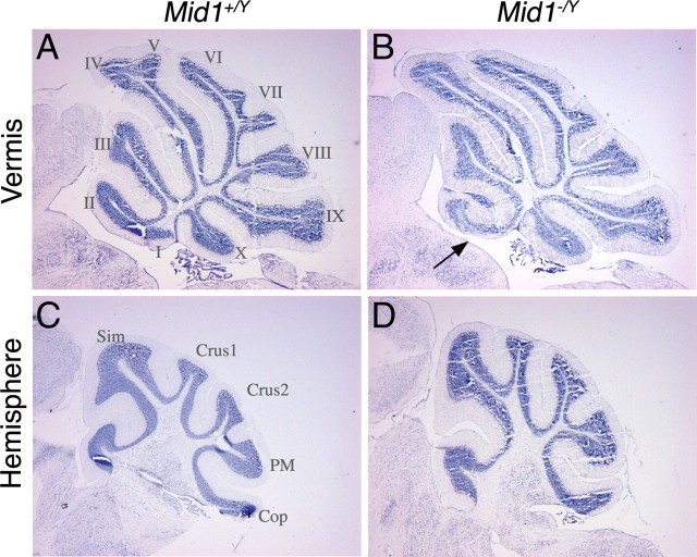
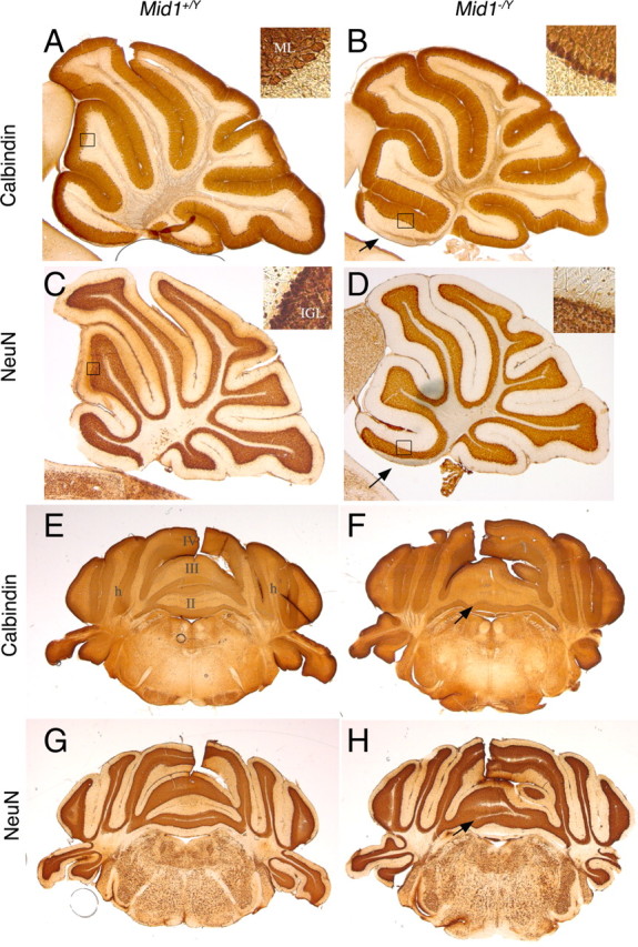
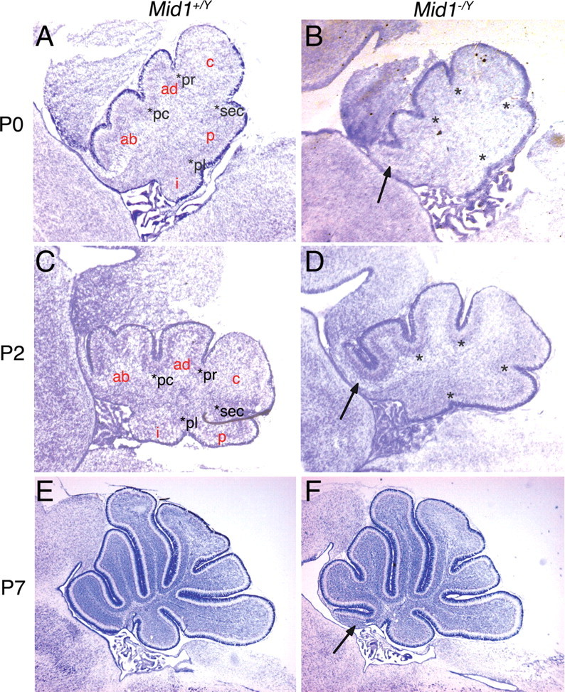
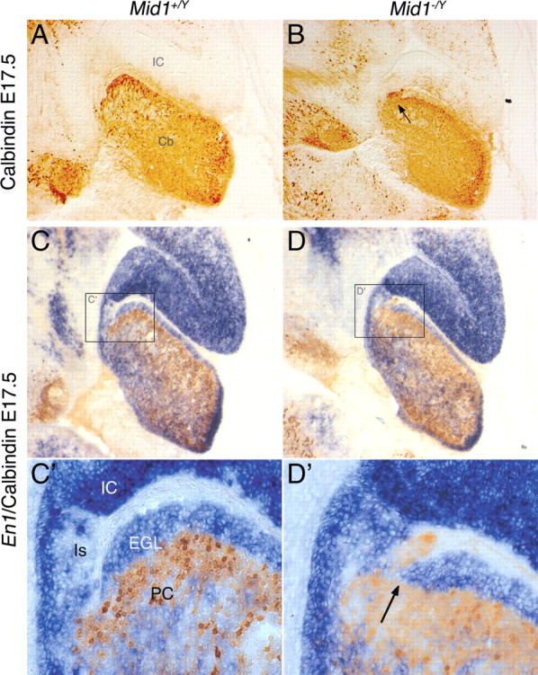
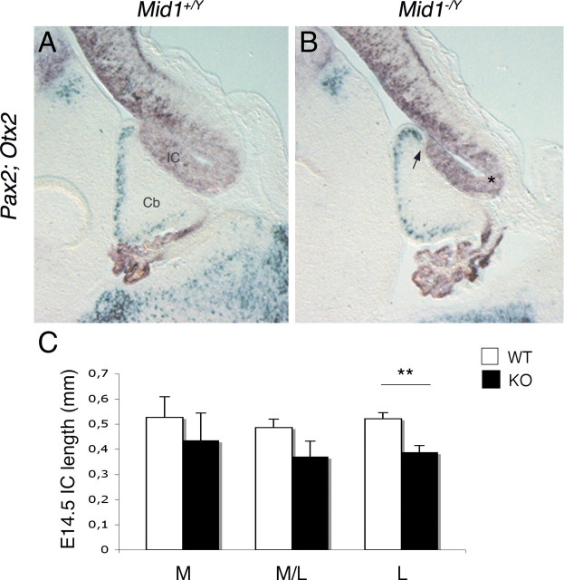
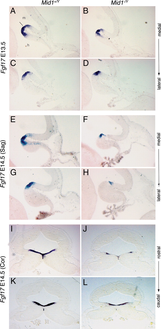
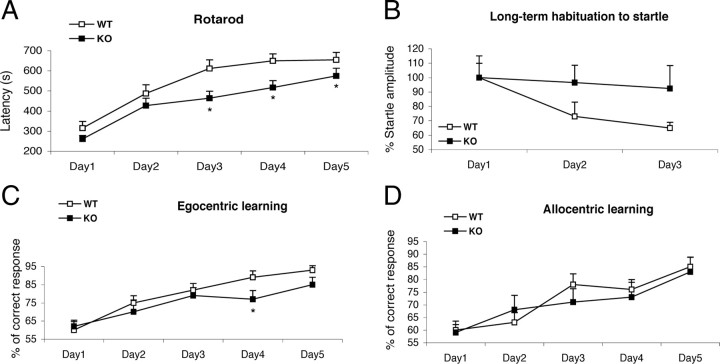
Opitz G/BBB syndrome (OS) is a genetic disorder characterized by midline developmental defects. Male patients with the X-linked form of OS, caused by loss-of-function mutations in the MID1 gene, show high variability of the clinical signs. MID1 encodes a ubiquitin ligase that controls phosphatase 2A, but its role in the pathogenesis of the disease is still unclear. Here, we report a mouse line carrying a nonfunctional ortholog of the human MID1 gene, Mid1. Mid1-null mice show the brain anatomical defect observed in patients (i.e., hypoplasia of the anterior portion of the medial cerebellum, the vermis). We found that the presence of this defect correlates with motor coordination and procedural and nonassociative learning impairments. The defect is limited to the most anterior lobes of the vermis, the region of the developing cerebellum adjacent to the dorsal midbrain. Analyses at midgestation reveal that lack of Mid1 causes the shortening of the posterior dorsal midbrain, the rostralization of the midbrain/cerebellum boundary, and the downregulation of a key player in the development of this region, Fgf17. Thus, lack of Mid1 causes a misspecification of the midbrain/cerebellar boundary that results in an abnormal development of the most anterior cerebellar lobes. This animal model provides a tool for additional in vivo studies of the physiological and pathological role of the Mid1 gene and a system to investigate the development and function of anterior cerebellar domains.


Figures

Figure 1.
Figure 1.
Abnormal cerebellum in Mid1 −/Y mice. Sagittal sections through the cerebellar vermis (A, B) and hemisphere (C, D) of adult wild-type and null mice stained with Nissl. Anterior is to the left. The numbers of the vermal and names of lateral lobes are indicated. The arrow in B indicates the anterobasal defect in Mid1 −/Y mice.
Figure 2.
Figure 2.
Normal ML and IGL organization in Mid1 −/Y adult mice. Sagittal sections through the vermis (A–D) and coronal sections at the level of the anterior cerebellum (E–H) are shown. The insets show magnification of the indicated areas. In null mice, PCs and their dendrites form a normal ML (detected by anti-Calbindin) and granule cells form a normal IGL (detected by anti-NeuN). The numbers of the vermal lobes are indicated in E. h, Hemisphere. The arrows indicate the defect in Mid1 −/Y brains.
Figure 3.
Figure 3.
The anterobasal lobe defect in Mid1 −/Y mice is detected at birth. Sagittal sections through the vermis at birth (A, B), P2 (C, D), and P7 (E, F) stained with Nissl. The granule cells at P0 and P2 are still superficial and form the EGL, whereas at P7 they are starting to migrate inwards to eventually form the IGL. The four principal fissures (pc, preculminate; pr, primary; sec, secondary; pl, posterolateral) are normally formed in P0 and P2 Mid1 −/Y mice. The cardinal lobes generated by the principal fissures are correctly formed with the exception of the anterobasal lobe, rostral to the preculminate fissure, that shows the defect (ab, anterobasal; ad, anterodorsal; c, central; p, posterior; i, inferior). The development of the other cardinal lobes into the definitive foliation proceeds normally from P0 through P7. The asterisks indicate the principal fissures. The arrows indicate the defect.
Figure 4.
Figure 4.
Inaccurate definition of the dorsal midbrain/cerebellum boundary in Mid1 −/Y mice. Immunohistochemistry with anti-Calbindin (A, B) and RNA in situ hybridization (En1) followed by anti-Calbindin (C–D′) on medial sagittal sections of E17.5 brains. C′, D′, Magnification of the delimited areas. Cb, Cerebellum; Is, isthmus. The arrows indicate ectopic PCs in Mid1 −/Y embryos.
Figure 5.
Figure 5.
Reduced IC length and rostralization of midbrain/cerebellar boundary in Mid1 −/Y embryos. Overlay of images of adjacent E14.5 sagittal sections hybridized with Pax2 (green) and Otx2 (purple) (A, B). The arrow indicates the mispositioning of the isthmus (B). C, E14.5 IC length (in millimeters) in wild-type (n = 4) and null (n = 4) mice. The length is the ventricular side distance between the two physical bending, indicated with the arrow and the asterisk in B, at three different positions of the vermal region: M, medial; M/L, mediolateral (∼120 μm from the midline); L, lateral (∼240 μm from the midline). t test, **p = 0.0042. Error bars indicate SEM.
Figure 6.
Figure 6.
Fgf17 is downregulated in Mid1 −/Y embryos. RNA in situ hybridization of Fgf17 on E13.5 sagittal section [medial (A, B); mediolateral (C, D)], on E14.5 sagittal sections [medial (E, F); mediolateral (G, H)], and on E14.5 coronal sections [rostral (I, J); caudal (K, L)]. Downregulation of Fgf17 in Mid1 −/Y embryos starts at E13.5 and is more evident at E14.5 especially in the next to the midline region along the mediolateral axis and in the cerebellum along the anteroposterior axis. The line indicates the isthmus. m, Midbrain; h, hindbrain.
Figure 7.
Figure 7.
Mid1 −/Y mice show motor coordination and learning impairments. A, Mid1 −/Y mice demonstrate a worse performance in the rotarod task, showing a deficit in motor learning acquisition (F ( 1 , 48 ) = 5.042; p = 0.02). B, Control mice (p = 0.008), but not Mid1 −/Y mice, reduce the percentage of startle amplitude across days in the long-term habituation of acoustic startle task; the null mice do not habituate to the stimulus; the startle response between the two groups significantly differ on the third day (p = 0.02). C, D, Mid1 −/Y mice recorded fewer correct responses in the egocentric spatial version of the cross maze task (C) on the fourth (p = 0.01) and on the fifth day (p = 0.06), but not in the allocentric one (D). *p < 0.05, Mid1 −/Y versus wild type. Error bars indicate SEM.

Similar articles

Cited by

References

    1. Biesecker LG. What you can learn from one gene: GLI3. J Med Genet. 2006;43:465–469. - PMC - PubMed
    1. Blaess S, Stephen D, Joyner AL. Gli3 coordinates three-dimensional patterning and growth of the tectum and cerebellum by integrating Shh and Fgf8 signaling. Development. 2008;135:2093–2103. - PMC - PubMed
    1. Bobée S, Mariette E, Tremblay-Leveau H, Caston J. Effects of early midline cerebellar lesion on cognitive and emotional functions in the rat. Behav Brain Res. 2000;112:107–117. - PubMed
    1. Broccoli V, Boncinelli E, Wurst W. The caudal limit of Otx2 expression positions the isthmic organizer. Nature. 1999;401:164–168. - PubMed
    1. Buchner G, Montini E, Andolfi G, Quaderi N, Cainarca S, Messali S, Bassi MT, Ballabio A, Meroni G, Franco B. MID2, a homologue of the Opitz syndrome gene MID1: similarities in subcellular localization and differences in expression during development. Hum Mol Genet. 1999;8:1397–1407. - PubMed

Publication types

MeSH terms

Grants and funding