. 2010 Feb 24;30(8):2897-910.
doi: 10.1523/JNEUROSCI.5725-09.2010.

The coxsackievirus-adenovirus receptor reveals complex homophilic and heterophilic interactions on neural cells

Affiliations

The coxsackievirus-adenovirus receptor reveals complex homophilic and heterophilic interactions on neural cells

Christopher Patzke et al. J Neurosci. .

Abstract

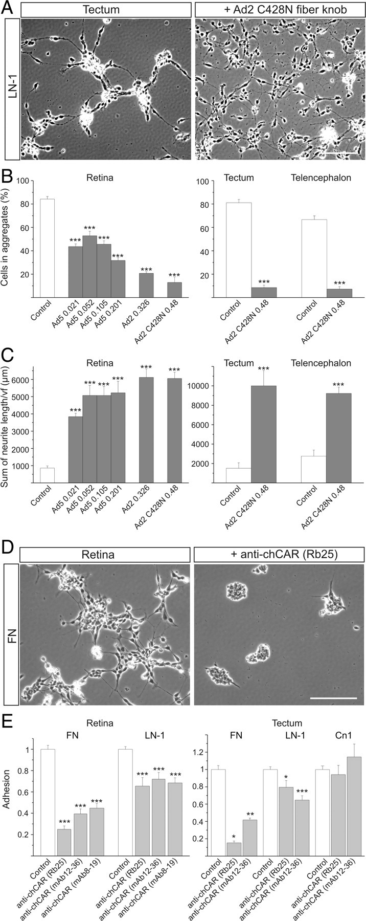
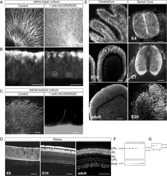
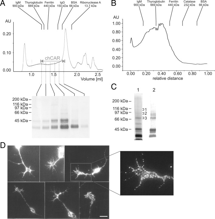
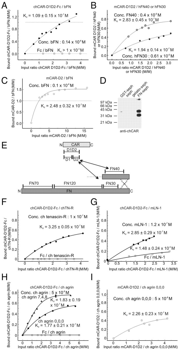
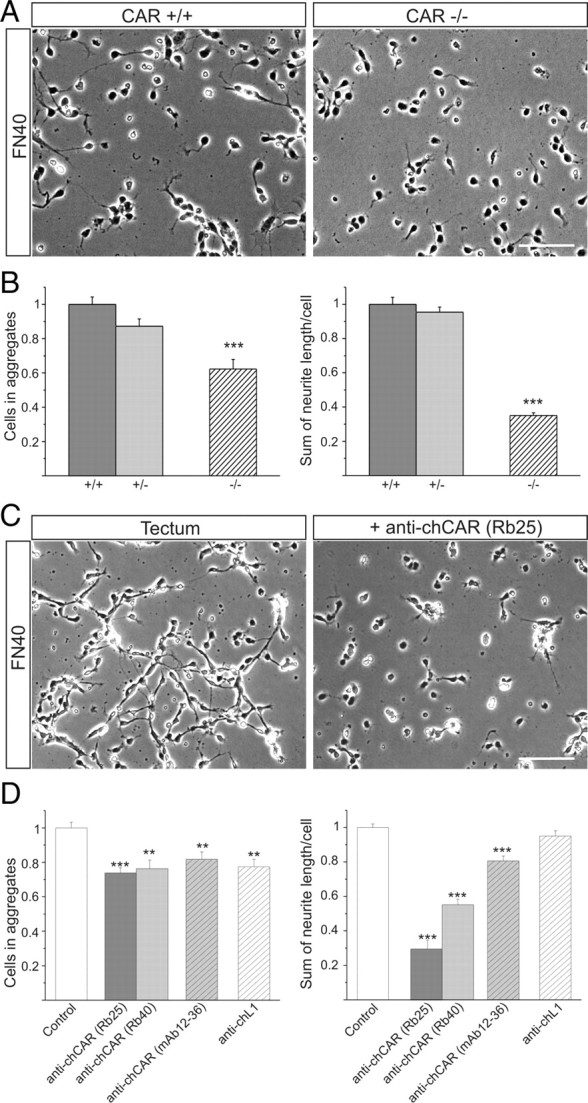
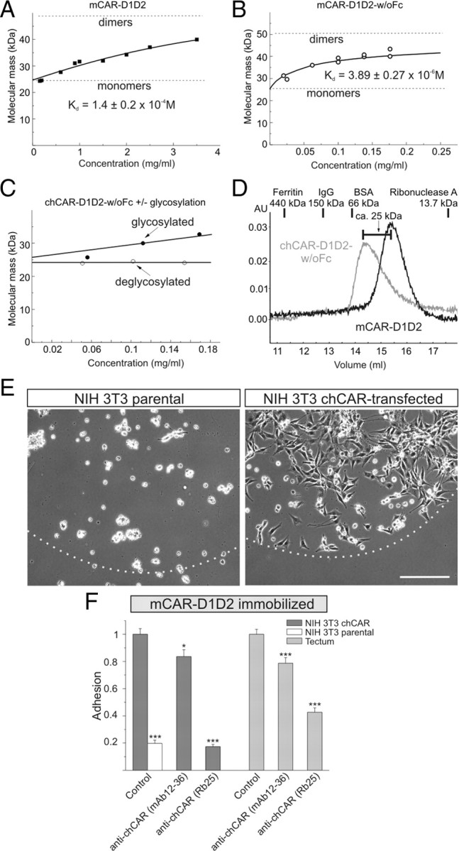
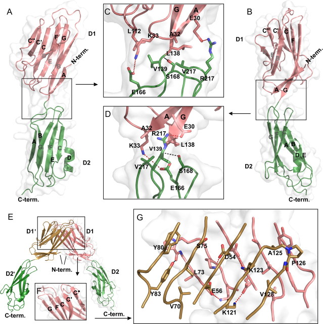
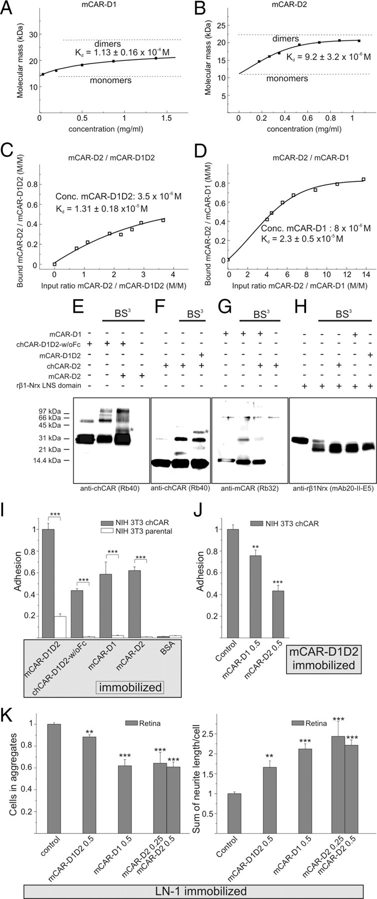
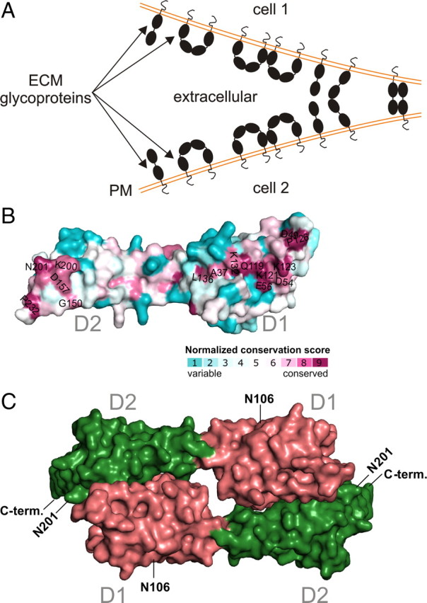
The coxsackievirus-adenovirus receptor (CAR) is a member of the Ig superfamily strongly expressed in the developing nervous system. Our histological investigations during development reveal an initial uniform distribution of CAR on all neural cells with a concentration on membranes that face the margins of the nervous system (e.g., the basal laminae and the ventricular side). At more advanced stages, CAR becomes downregulated and restricted to specific regions including areas rich in axonal and dendritic surfaces. To study the function of CAR on neural cells, we used the fiber knob of the adenovirus, extracellular CAR domains, blocking antibodies to CAR, as well as CAR-deficient neural cells. Blocking antibodies were found to inhibit neurite extension in retina organ and retinal explant cultures, whereas the application of the recombinant fiber knob of the adenovirus subtype Ad2 or extracellular CAR domains promoted neurite extension and adhesion to extracellular matrices. We observed a promiscuous interaction of CAR with extracellular matrix glycoproteins, which was deduced from analytical ultracentrifugation experiments, affinity chromatography, and adhesion assays. The membrane proximal Ig domain of CAR, termed D2, was found to bind to a fibronectin fragment, including the heparin-binding domain 2, which promotes neurite extension of wild type, but not of CAR-deficient neural cells. In contrast to heterophilic interactions, homophilic association of CAR involves both Ig domains, as was revealed by ultracentrifugation, chemical cross-linking, and adhesion studies. The results of these functional and binding studies are correlated to a U-shaped homodimer of the complete extracellular domains of CAR detected by x-ray crystallography.


Figures

Figure 1.
Figure 1.
Attachment and neurite extension to ECM glycoproteins in the presence of the fiber knob of the adenovirus or anti-CAR antibodies. A–C, In the presence of the fiber knob, the number of single neural cells from chicken embryos (E8) attached and the total length of measurable neurites increased on immobilized LN-1. Clusters of somata of more than five cells were considered as aggregates. Concentrations of fiber knobs (Ad2, Ad2C428N, Ad5) from different adenoviruses are indicated in milligrams per milliliter. D, E, Attachment of retinal cells to FN or LN-1 is reduced in the presence of antibodies to CAR. The mean number of attached cells in the presence of antibodies to CAR is compared with untreated cells. Polyclonal rabbit antibodies (Rb25) were applied in the form of Fab fragments (0.25 mg/ml) and mAbs as intact IgGs (10 μg/ml). The specificity of the polyclonal antibodies were tested by preincubation with affinity-purified CAR, which resulted in complete neutralization of the antibody-mediated effect. Scale bars, 100 μm. Error bars indicate SEM. *p < 0.05; **p < 0.005; ***p < 0.0005.
Figure 2.
Figure 2.
The outgrowth of RGC axons in retina organ cultures and on basal laminae is reduced in the presence of antibodies to chCAR. A, RGC axons in flat-mounted retina organ cultures in the presence of Fab fragments of polyclonal antibodies to chCAR or control antibodies (0.5 mg/ml). Chick embryonic eyes (E4.5) were cultivated after removal of the pigment epithelium for 20 h. After fixation, retinae were flat mounted, and RGC axons were visualized by anti-chL1 staining. B, Cross sections of retina organ cultures. In the developing optic fiber layer (top), fewer axon bundles of RGCs are observed in the presence of Fab fragments of anti-CAR antibodies. C, E6 retinal explants incubated for 24 h on basal laminae preparations from chick retinae in the presence of Fab fragments of polyclonal antibodies to chCAR (0.5 mg/ml) or control antibodies. D, Localization of CAR in the developing chick retina. Cryostat sections of E6, E10, or adult retinae were stained indirectly by mAb12-36 to chCAR. OFL, Optic fiber layer; IPL, inner plexiform layer; INL, inner nuclear layer; OPL, outer plexiform layer; PhR, photoreceptor layer. E, Localization of CAR in cryostat sections of chicken cerebellum or spinal cord at different developmental stages using mAb12-36. The E20 spinal cord section shows only the dorsolateral half of the cord. F, Western blot analysis of the chick retinae from different developmental stages (E7–E20) using mAb12-36. Note that in contrast to CAR, Cn1 is upregulated during development. Lanes were loaded with equal amounts of protein (5 μg). G, A developmental gradient of CAR within the retina is revealed by Western blotting. The E16 retina was cut into three parts: central (C), intermediate (I), or peripheral (P) retina. Equal amounts of protein from each part was loaded on SDS-PAGE and analyzed in Western blots using mAb12-36. Scale bars, 100 μm.
Figure 3.
Figure 3.
CAR exists in protein complexes on neural surfaces. A–C, CAR from chicken brain exists in complexes as analyzed by gel filtration or BN gels. Detergent (1% CHAPS) extracts of plasma membrane preparations from embryonic chicken brains were run over a Superose 6 PC column, and CAR components were detected in Western blotting by Rb54. BN gels containing 4–12% acrylamide were blotted, and a densitometric scan of the Western blot using Rb54 is shown. [Note that CAR purified from detergent extracts by immunoaffinity or gel filtration chromatography consists of two major components at 36 and 46 kDa. The 36 kDa component contains the N terminus of the mature CAR polypeptide as determined by Edmann degradation (LSITSAESAFEKAQGER), suggesting that it results from C-terminal degradations of CAR.] Molecular masses of standard proteins are given at the top or on the left of each panel. C, chCAR obtained by immunoaffinity chromatography from detergent extracts of plasma membrane preparations of embryonic chicken brains reveals SDS-resistant complexes in SDS-PAGE under reducing conditions as visualized by silver staining (lane 1). Lane 2 shows a Western blot analysis of the affinity isolate as revealed in lane 1 using Rb54 to chCAR. In addition to the 36 and 46 kDa bands, minor components at 72, 95, and 110 kDa were also detected. The identity of these proteins was further established by mass spectrometry sequencing of a tryptic digest that yielded chCAR peptides encompassing the following amino acid residues (the position are given): 72 kDa (3): 71–82, 72–82, 83–94, 95–103, 104–115, 128–136, 150–160, 182–197, 210–222, 305–320, 321–333, 354–366; 95 kDa (2): 71–82, 83–94, 95–103, 104–115, 128–136, 137–149, 166–180, 181–197, 182–192, 210–222, 277–290, 305–320, 321–333, 354–366; 110 kDa (1): 71–82, 72–82, 95–103, 104–115, 128–136, 166–180, 182–197, 210–222, 305–320, 321–333, 354–366. D, Localization of CAR on chick tectal neurons cultivated on LN-1. CAR was stained after fixation using mAb12-36. One growth cone is enlarged on the right. Scale bar, 20 μm. AU, Arbitrary units.
Figure 4.
Figure 4.
D2 of CAR binds to FN, FN30, or FN40, and CAR interacts with the ECM glycoprotein TN-R, LN-1, or agrin. A, Equilibrium sedimentation of chCAR-D1D2-Fc and bFN. Note that at a 3.5-fold molar excess of chCAR-D1D2-Fc, more than one chCAR-D1D2-Fc is bound to FN. The Fc fragment served as the control. A concentration of 0.14 μm chCAR-D1D2-Fc corresponds to 16.9 μg/ml. B, mCAR-D1D2 binds to the fragment FN40 or FN30 (see also supplemental Fig. S3, available at www.jneurosci.org as supplemental material). A concentration of 0.4 μm mCAR-D1D2 corresponds to 9.6 μg/ml. C, Interaction of mCAR-D2 and FN is demonstrated by equilibrium sedimentation. A concentration of 0.1 μm mCAR-D2 corresponds to 1.2 μg/ml. D, chCAR enriched on a FN-Sepharose (seph) column. Equal volumes of detergent extracts of plasma membrane preparation from E15 chicken brains were passed over different affinity columns, washed, and eluted by diethylamine at pH 11.5, followed by Western blotting. chCAR appears under nonreducing conditions as bands of 42 and 32 kDa (see also Fig. 3C). E, Scheme of the CAR–FN interaction. Locations of the FN fragments and the N or C termini are indicated. Only a part of the two polypeptides of a FN dimer is shown. F–I, Equilibrium sedimentation analyses of interactions between chCAR-D1D2-Fc and TN-R, LN-1, and of the splice variants of agrin 0,0,0 or 7,4,8, and between mCAR-D1D2 and agrin 0,0,0. The Fc fragment served as the control. Conc., Concentration.
Figure 5.
Figure 5.
The CAR–FN40 interaction is important for neurite extension. A, B, E11 wild-type or CAR-deficient neural cells were plated on immobilized FN40. CAR−/− cells extend fewer neurites and form fewer aggregates compared with CAR+/+ cells. C, D, Aggregate formation and neurite extension of chick tectal cells (E6) on FN40 is blocked by antibodies to chCAR, whereas antibodies to chL1 do not interfere with neurite extension. Clusters of more than three cells were considered as aggregates. Cell numbers are expressed as mean per optical view field ± SEM and were normalized to wild-type or untreated cells. Neurite length was determined per view field divided by number of attached cells. **p < 0.005; ***p < 0.0005. Scale bars, 100 μm.
Figure 6.
Figure 6.
Homophilic binding of CAR-D1D2 domains is enhanced by N-glycosylation, and CAR-expressing cells bind to immobilized mCAR-D1D2. A, Equilibrium sedimentation analysis of mCAR-D1D2. At concentrations up to 3.5 mg/ml, a monomer–dimer equilibrium is observed. B, mCAR-D1D2-w/oFc forms a monomer–dimer equilibrium. Note that in contrast to mCAR-D1D2, 50% of mCAR-D1D2-w/oFc are found in dimers at a concentration of 170 μg/ml. C, At concentrations up to 170 μg/ml, self-association of chCAR-D1D2-w/oFc is enhanced by N-glycosylation. chCAR-D1D2-w/oFc was deglycosylated by PNGaseF for 2 h at 37°C. Self-association was monitored at three different concentrations by equilibrium sedimentation. Underglycosylated chCAR-D1D2-w/oFc served as the control. D, Comparison of size exclusion chromatography profiles (Superdex 200 HR) of mCAR-D1D2 (black) and chCAR-D1D2-w/oFc (gray). Identical concentrations were applied (0.5 ml of 130 μg/ml at a flow rate of 0.5 ml/min in PBS). mCAR-D1D2 runs predominantly as a monomer, and chCAR-D1D2-w/oFc runs predominantly as a dimer. The positions of standard proteins are shown at the top. The calculated size difference between the peaks of mCAR-D1D2 and chCAR-D1D2-w/oFc is equivalent to the size of the mCAR-D1D2 monomer (25 kDa). The theoretical masses are 24378.5 and 24433.6 for chCAR-D1D2-w/oFc and mCAR-D1D2, respectively, and the measured mass of glycosylated chCAR-D1D2-w/oFc is 26081.5 as determined by mass spectrometry. E, F, chCAR-transfected NIH 3T3 cells attach to immobilized mCAR-D1D2 and spread while parental cells are unable to attach. Attachment is disturbed by species-specific antibodies to chCAR (Rb25 or mAb12–36). Culture dishes were precoated with 2 μl of mCAR-D1D2 (100 μg/ml) in their center. The border of the coated area is indicated by a broken line. Means ± SEM are normalized. *p < 0.05; ***p < 0.0005. Scale bar, 200 μm.
Figure 7.
Figure 7.
Crystal structure of extracellular mCAR-D1D2 reveals a U-shaped dimer. A–D, Secondary and tertiary structure of mCAR-D1D2; parts belonging to D1 and D2 are colored pink and green, respectively. The molecular contact area is displayed as a transparent gray surface. A, B, Overview of the Ig folds in D1 and D2. β-Strands in D1 and D2 are labeled in uppercase letters. Labels of the two sheets forming a β-sandwich are colored black and gray, respectively. The view in B is rotated by 90° with respect to A. C, D, Layout of the D1–D2 junction. Side chains defining the junction are displayed as sticks, with noncarbon atoms colored according to CPK conventions. Hydrogen bonds and salt bridges/electrostatic contacts are shown as dashed black and red lines, respectively. The view in D is rotated by 90° with respect to A. E, Overview of two symmetry-related mCAR monomers (D1 and D2 vs D1′ and D2′) forming a U-shaped dimer. The contact surface is shown only for one mCAR molecule (D1 and D2), and the D1′ domain is colored brown. F, Structural elements forming the dimer interface shown for D1 in E. G, Detailed view of interactions inside the dimer interface shown in E. The layout according to C and D is shown. For a better overview, labels are shown for domain D1′ only but can be inferred to D1 by symmetry. C-term., C terminus; N-term., N terminus.
Figure 8.
Figure 8.
Homophilic adhesion is mediated by D1 and D2. A–D, mCAR-D1 and mCAR-D2 are able to self-associate as revealed by analytical ultracentrifugation and represent a monomer–dimer equilibrium in solution. mCAR-D1 or mCAR-D2 binds to mCAR-D1D2. E–H, Chemical cross-linking of CAR polypeptides. Western blots of extracellular CAR domains probed with antibodies against chCAR or mCAR are shown. E, chCAR-D1D2-w/oFc migrates as a band at 30 kDa and a weaker band at 60 kDa, which represents a dimer. Cross-linking with BS3 leads to an increase of dimers and to the occurrence of higher oligomers as well, and to a band at 40 kDa, which represents a heterodimer of chCAR-D1D2-w/oFc and mCAR-D2. F, Similar results are revealed when mCAR-D1D2 and chCAR-D2 are cross-linked. (mCAR-D1D2 migrates as a band of 25 kDa and a weak dimer band at 50 kDa.) G, chCAR-D2 and mCAR-D1 migrate as a monomer and dimer. (Note that the dimeric mCAR-D1 band at 30 kDa becomes less intense because of the binding chD2 that is not recognized by anti-mouse CAR.) H, mCAR domains are not cross-linked to the β1-LNS domain of rat neurexin. I, chCAR-expressing 3T3 cells adhered to immobilized mCAR-D1D2, chCAR-D1D2-w/oFc, mCAR-D1, or mCAR-D2. J, The attachment of CAR-expressing 3T3 cells was blocked by mCAR-D1 or mCAR-D2 in solution at a concentration of 0.5 mg/ml. K, Formation of aggregates of tectal cells was blocked by mCAR-D1 or mCAR-D2 in solution while the formation of neurites was promoted. Concentrations (Conc.) are indicated in milligrams per milliliters. Error bars indicate SEM. **p < 0.005; ***p < 0.0005.
Figure 9.
Figure 9.
Summary of molecular interactions of CAR. A, Scheme of putative molecular interactions of CAR on the neural plasma membrane (PM). Possible homophilic interactions of CAR are as follows: the D1–D1 self-association revealed by the U-shaped crystal structure most likely occurs between CAR polypeptides attached to the same plasma membrane. Additional binding and adhesion data suggest that homophilic interactions of CAR between two cells result from an antiparallel D1–D2 interaction. Heterophilic interactions to ECM glycoproteins are indicated by arrows. B, C, Proposed model for two mCAR-D1D2 monomers associated via two D1–D2 interfaces based on molecular docking simulations. Molecular contact surfaces corresponding to D1 and D2 are colored pink and green, respectively. Glycosylation sites (N106 and N201) and C termini are labeled. Normalized conservation score indicated by a color code. . C-term., C terminus; N-term., N terminus.

References

    1. Altschul SF, Gish W, Miller W, Myers EW, Lipman DJ. Basic local alignment search tool. J Mol Biol. 1990;215:403–410. - PubMed
    1. Asher DR, Cerny AM, Weiler SR, Horner JW, Keeler ML, Neptune MA, Jones SN, Bronson RT, DePinho RA, Finberg RW. Coxsackievirus and adenovirus receptor is essential for cardiomyocyte development. Genesis. 2005;42:77–85. - PubMed
    1. Awasthi V, Meinken G, Springer K, Srivastava SC, Freimuth P. Biodistribution of radioiodinated adenovirus fiber protein knob domain after intravenous injection in mice. J Virol. 2004;78:6431–6438. - PMC - PubMed
    1. Bailey S. The Ccp4 Suite—Programs for Protein Crystallography. Acta Crystallogr D Biol Crystallogr. 1994;50:760–763. - PubMed
    1. Behlke J, Ristau O, Schonfeld HJ. Nucleotide-dependent complex formation between the Escherichia coli chaperonins GroEL and GroES studied under equilibrium conditions. Biochemistry. 1997;36:5149–5156. - PubMed

Publication types

MeSH terms

LinkOut - more resources