. 2010 Feb 24;30(8):2935-50.
doi: 10.1523/JNEUROSCI.4411-09.2010.

Dopamine-dependent tuning of striatal inhibitory synaptogenesis

Affiliations

Dopamine-dependent tuning of striatal inhibitory synaptogenesis

Darren Goffin et al. J Neurosci. .

Abstract

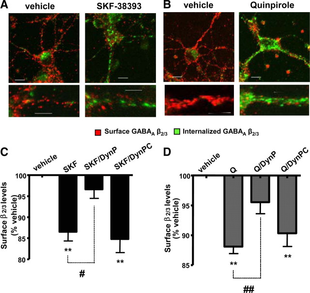
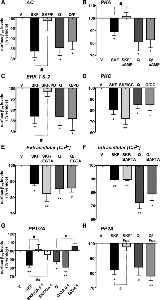
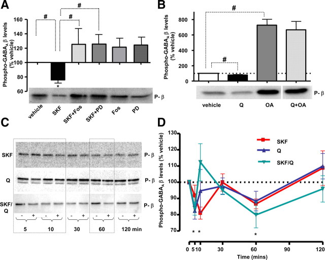
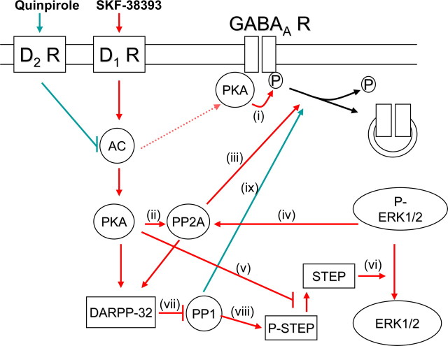
Dopaminergic projections to the striatum, crucial for the correct functioning of this brain region in adulthood, are known to be established early in development, but their role is currently uncharacterized. We demonstrate here that dopamine, by activating D(1)- and/or D(2)-dopamine receptors, decreases the number of functional GABAergic synapses formed between the embryonic precursors of the medium spiny neurons, the principal output neurons of the striatum, with associated changes in spontaneous synaptic activity. Activation of these receptors reduces the size of postsynaptic GABA(A) receptor clusters and their overall cell-surface expression, without affecting the total number of clusters or the size or number of GABAergic nerve terminals. These changes result from an increased internalization of GABA(A) receptors, and are mediated by distinct signaling pathways converging at the level of GABA(A) receptors to cause a transient PP2A/PP1-dependent dephosphorylation. Thus, tonic D(1)- and D(2)-receptor activity limits the extent of collateral inhibitory synaptogenesis between medium spiny neurons, revealing a novel role of dopamine in controlling the development of intrinsic striatal microcircuits.


Figures

Figure 1.
Figure 1.
Dopaminergic projections and dopamine receptors are present in the embryonic striatum. A, Dopaminergic projections positive for TH (×5, scale bar corresponds to 200 μm) form a prominent network of fibers in the ventral part of the striatal enlargement, while terminal-like staining (×63, scale bars correspond to 20 μm) is abundant in the dorsal part of the embryonic (E17) striatum. B, Dopamine D1 receptors and D2 receptors are expressed throughout the whole striatal enlargement and show a high-degree of colocalization (×63, scale bars correspond to 20 μm).
Figure 2.
Figure 2.
Dopamine receptor activation decreases the colocalization of postsynaptic GABAA receptor β2/3 subunit puncta with presynaptic VIAAT-1 puncta. Embryonic striatal neurons express both D1Rs and D2Rs when cultured for 7 d (7 DIV) (A), and 14 d (14 DIV) (B). Scale bars correspond to 20 μm. Striatal cultures were treated for 72 h (from 4 to 7 DIV) (C) or 7 d (from 7 to 14 DIV) (D) with vehicle, SKF-38393, or quinpirole, and stained for GABAAR β2/3 subunit (in red) and VIAAT-1 (in green). Scale bars correspond to 10 μm. The percentage of β2/3 subunit-positive puncta that colocalized with VIAAT-1 puncta was significantly decreased in cultures treated with SKF-38393 or quinpirole for 72 h (E) or 7 d (F). G, H, Relative cumulative frequency plots of β2/3-immunoreactive puncta and VIAAT-1-positive puncta (in square micrometers) measured along a defined length of primary processes (20 μm) following the treatment with vehicle (white), SKF-38393 (black), or quinpirole (dark gray) for 72 h (4–7 DIV, left) (G) or 7 d (7–14 DIV, right) (H). I, SKF-38393 (black) and quinpirole (gray) significantly decreased the average β2/3 subunit punctum area in comparison with controls (vehicle treated, white) cells, following treatments for 72 h or 7 d. J, SKF-38393 (black) and quinpirole (gray) had no effect on the average VIAAT-1 punctum area in comparison with controls (vehicle treated, white), following treatments for 72 h or 7 d. K, The total number of β2/3-positive puncta remained unchanged following the treatment with vehicle (white), SKF-38393 (black), or quinpirole (gray) for 72 h or 7 d. L, The total number of VIAAT-1-positive puncta remained unchanged following the treatment with vehicle (white), SKF-38393 (black), or quinpirole (gray) for 72 h or 7 d. Bars represent mean ± SEM. Statistical analysis was performed using paired t test: ***p < 0.001.
Figure 3.
Figure 3.
Dopamine receptor activation decreases the number of functional GABAergic synapses. A, Cultures were treated for 72 h (from 4 to 7 DIV) with vehicle, SKF-38393, or quinpirole, followed by activity-dependent labeling with FM1-43FX. Scale bars correspond to 10 μm. SKF-38393 and quinpirole significantly decreased the colocalization of β2/3 puncta (in red) and FM1-43FX puncta (in green) after treatment for 72 h (from 4 to 7 DIV, B), or 7 d (from 7 to 14 DIV, C). Bars represent mean ± SEM. Statistics was performed using t test: ***p < 0.001. D, mIPSPs recorded in striatal neurons after 72 h treatment (from 4 to 7 DIV) with vehicle (control) or SKF-38393 in the presence of TTX (1 μm), d-AP5 (50 mm), and CNQX (20 mm), before and after bath application of GABAA receptor antagonist picrotoxin (50 μm, + picrotoxin). Three representative traces for each condition are shown. Scale refers to all four conditions. E, F, SKF-38393 and quinpirole treatments decreased the amplitude (E) and frequency (F) of mIPSPs. Bars represent mean ± SD. Statistical analysis was performed using two-tailed t test: *p < 0.05.
Figure 4.
Figure 4.
Activation of dopamine receptors decreases the cell-surface expression of GABAA receptors. A, SKF-38393 (1 nm) caused a significant and prolonged decrease in the level of β2/3 subunits (black) expressed at the cell surface, but not in total levels of these subunits (gray). B, Quinpirole (100 nm) caused a significant and prolonged decrease in β2/3 subunit cell-surface expression (black) but not in their total levels (gray). C, D1R antagonist SCH-23390 (1 μm, SCH) abolished the reduction in surface β2/3 levels upon 30 min treatment with SKF-38393 (1 nm, SKF). D, D2R antagonist sulpiride (10 μm, Sulp) significantly attenuated the reduction in surface β2/3 levels upon 30 min treatment with quinpirole (100 nm, Q). E, Dopamine (1 μm, DA)-dependent decrease in the β2/3 subunit surface was significantly attenuated by SCH-23390 (DA + SCH), and sulpiride (DA + Sulp), indicating that both D1Rs and D2Rs mediate the effects of dopamine. F, Activation of both D1Rs and D2Rs by their respective agonists (SKF + Q) caused a reduction in surface β2/3 levels comparable to changes observed upon the treatment with SKF-38393 (SKF) or quinpirole (Q) alone. Bars represent mean ± SEM. Statistical analysis was performed using one-way ANOVA with either a Dunnett post hoc analysis versus control (*p < 0.05; **p < 0.01), or paired t test (#p < 0.05).
Figure 5.
Figure 5.
Dopamine receptor activation decreases the cell-surface expression of GABAA receptors via a dynamin-dependent pathway. A, B, Internalization of GABAA receptors was visualized by staining with β2/3-specific antibody in the presence of vehicle, SKF-38393 (A), or quinpirole (B), as described in Materials and Methods. Binding of β2/3-specific antibody to the surface expressed GABAA receptors was visualized using a secondary anti-mouse IgG coupled to Alexa555 (in red), while binding of the same antibody to internalized receptors was visualized using a secondary anti-mouse IgG coupled to Alexa488 (in green). Scale bars correspond to 10 μm. C, The decrease in surface β2/3 subunit levels caused by SKF-38393 (SKF) was significantly attenuated in the presence of a dynamin inhibitory peptide (SKF/DynP), but not in the presence of its scrambled control (SKF/DynPC). D, The decrease in surface β2/3 subunit levels caused by quinpirole (Q) was also significantly attenuated in the presence of a dynamin inhibitory peptide (Q/DynP), but not by its scrambled control (Q/DynPC). Bars represent mean ± SEM. Statistical analysis was performed using one-way ANOVA with Dunnett post hoc analysis versus control (*p < 0.05; **p < 0.01) or paired t test (#p < 0.05; ##p < 0.01).
Figure 6.
Figure 6.
D1R-dependent reduction of the GABAA receptor levels expressed at the cell surface is mediated by the activities of PKA, ERK1 and ERK2, and PP2A, whereas D2R-dependent decrease is mediated by the activity of PP1. A, The effect of SKF-38393 (SKF), but not quinpirole (Q), was occluded by a direct activation of adenylyl cyclase (AC) with forskolin (SKF/F, Q/F, respectively). B, The effect of SKF-38393 (SKF), but not quinpirole (Q), was also occluded by a direct activation of protein kinase A (PKA) with 8-bromo-cAMP (SKF/cAMP, Q/cAMP, respectively). C, The effect of SKF-38393 (SKF), but not quinpirole (Q) was attenuated by inhibition of ERK kinases (ERK1 & 2) with PD-98059 (SKF/PD, Q/PD, respectively). D, The inhibition of protein kinase C (PKC) by calphostin C (CC) had no effect on SKF-38393-dependent (SKF vs SKF/CC), or quinpirole-dependent (Q vs Q/CC) decrease in β2/3 surface levels. E, Buffering of extracellular calcium with EGTA (EGTA) had no effect on SKF-38393-dependent (SKF vs SKF/EGTA), or quinpirole-dependent (Q vs Q/EGTA) decrease in β2/3 surface levels. F, Buffering of intracellular calcium with BAPTA-AM (BAPTA) had no effect on SKF-38393-dependent (SKF vs SKF/BAPTA), or quinpirole-dependent (Q vs Q/BAPTA) decrease in β2/3 surface levels. G, The decrease in surface β2/3 levels caused by SKF-38393 (SKF), but not quinpirole (Q) was abolished by a low concentration of okadaic acid (0.1 μm; SKF/OA 0.1, Q/OA 0.1, respectively), while both SKF-38393-dependent (SKF) and quinpirole-dependent (Q) decreases were abolished by a high concentration of okadaic acid (1 μm; SKF/OA 1, Q/OA 1, respectively). H, The effect of SKF-38393 (SKF), but not quinpirole (Q), was significantly attenuated by fostriecin (SKF/Fos, Q/Fos, respectively). Bars represent mean ± SEM. Statistical analysis was performed using one-way ANOVA with either a Dunnett post hoc analysis versus control (*p < 0.05; **p < 0.01) or paired t test (#p < 0.05; ##p < 0.01).
Figure 7.
Figure 7.
D1R and D2R activation leads to a transient dephosphorylation of GABAA receptor β subunits. A, Treatment with SKF-38393 (1 nm, 30 min) led to a decrease in β subunit phosphorylation (SKF), which was abolished by fostriecin (SKF/Fos) or PD-98059 (SKF/PD). Both inhibitors alone caused small but insignificant increases in the basal phosphorylation state of the β subunits (Fos, PD, respectively). A representative immunoblot is shown below the bar graph. B, Treatment with quinpirole (100 nm, 30 min) also led to a decrease in β subunit phosphorylation (Q), which was abolished by okadaic acid (Q/OA). Okadaic acid alone caused a prominent increase in the basal phosphorylation state of the β subunits (OA). A representative immunoblot is shown below the bar graph. C, D, The time course of changes in the phosphorylation state of the β subunits in the presence of SKF-38393 (SKF), quinpirole (Q), or both (SKF/Q). Representative immunoblots (C) and graphs showing percentage values normalized to the appropriate controls treated with the vehicle (100%; D) show a transient decrease in the phosphorylation state of the β subunits at an early time point (5–10 min) and a later time point (60 min) of incubation with either SKF-38393 (SKF, in red), quinpirole (Q, in green), or both (SKF/Q, in blue). Values represent mean ± SEM. Statistical analysis was performed using one-way ANOVA with either a Dunnett post hoc analysis versus control (*p < 0.05) or paired t test (#p < 0.05).
Figure 8.
Figure 8.
Functional cross talk between D1R and D2R signaling pathways regulating GABAA receptors in the embryonic striatal neurons. Activation of D1R with SKF-38393 (in red) leads to activation of adenylyl cyclase (AC) resulting in activation of conceivably two segregated pools of protein kinase A (PKA). The first pool of PKA is associated with GABAA receptors leading to a rapid phosphorylation of GABAA receptors (i). The second pool of PKA is more distal and unable to associate with GABAA receptors. This pool of PKA directly phosphorylates and activates protein phosphatase 2A (PP2A, ii). Activated PP2A associates with and dephosphorylates GABAA receptors, promoting their internalization from the cell surface (iii). The ability of PP2A to dephosphorylate GABAA receptors is likely to be enhanced due to ERK1/2-mediated phosphorylation and activation of PP2A (iv). ERK1/2 can be activated by two different signaling pathways both involving PKA. First, PKA can directly phosphorylate and thus inactivate striatal enriched phosphatase (STEP, v). In this way, STEP is no longer able to dephosphorylate and deactivate ERK1/2. Second, PKA can lead to the activation of DARPP-32, an inhibitor of protein phosphatase 1 (PP1, vii). Inactivated PP1 cannot dephosphorylate and activate STEP, which is then unable to dephosphorylate ERK1/2 (viii). Both of these pathways lead to an increase in the level of phosphorylated ERK1/2, which acts on PP2A to promote dephosphorylation of GABAA receptors. Activation of D2Rs by quinpirole (in green) leads to a dephosphorylation of GABAA receptors and their internalization by inhibiting AC. Inhibition of AC leads to a reduction in the activation of PKA and consequently to a reduction in PKA-mediated phosphorylation of GABAA receptors (i). At the same time however, inhibition of AC may lead to a decrease in DARPP-32 activity and thus activation of PP1 (vii). Activated PP1 is then capable of associating with GABAA receptors and mediating their dephosphorylation and subsequent internalization (ix).

Similar articles

Cited by

References

    1. Ahn JH, McAvoy T, Rakhilin SV, Nishi A, Greengard P, Nairn AC. Protein kinase A activates protein phosphatase 2A by phosphorylation of the B56delta subunit. Proc Natl Acad Sci U S A. 2007;104:2979–2984. - PMC - PubMed
    1. Aizman O, Brismar H, Uhlén P, Zettergren E, Levey AI, Forssberg H, Greengard P, Aperia A. Anatomical and physiological evidence for D1 and D2 dopamine receptor colocalization in neostriatal neurons. Nat Neurosci. 2000;3:226–230. - PubMed
    1. Aosaki T, Kiuchi K, Kawaguchi Y. Dopamine D1-like receptor activation excites rat striatal large aspiny neurons in vitro. J Neurosci. 1998;18:5180–5190. - PMC - PubMed
    1. Awad JA, Johnson RA, Jakobs KH, Schultz G. Interactions of forskolin and adenylate cyclase. Effects on substrate kinetics and protection against inactivation by heat and N-ethylmaleimide. J Biol Chem. 1983;258:2960–2965. - PubMed
    1. Bedford FK, Kittler JT, Muller E, Thomas P, Uren JM, Merlo D, Wisden W, Triller A, Smart TG, Moss SJ. GABA(A) receptor cell surface number and subunit stability are regulated by the ubiquitin-like protein Plic-1. Nat Neurosci. 2001;4:908–916. - PubMed

Publication types

MeSH terms

LinkOut - more resources