. 2010 Feb 24;30(8):3002-12.
doi: 10.1523/JNEUROSCI.4851-09.2010.

Novel embryonic neuronal migration and proliferation defects in Dcx mutant mice are exacerbated by Lis1 reduction

Affiliations

Novel embryonic neuronal migration and proliferation defects in Dcx mutant mice are exacerbated by Lis1 reduction

Tiziano Pramparo et al. J Neurosci. .

Abstract

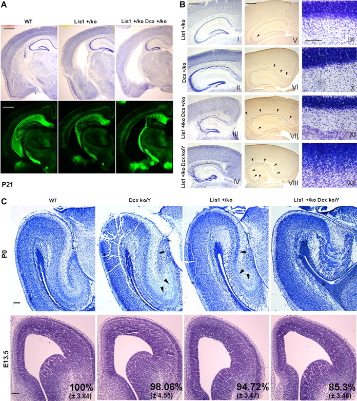
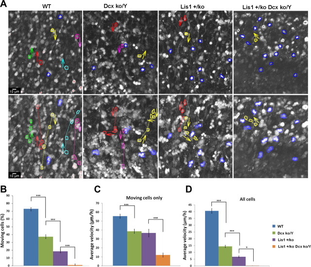
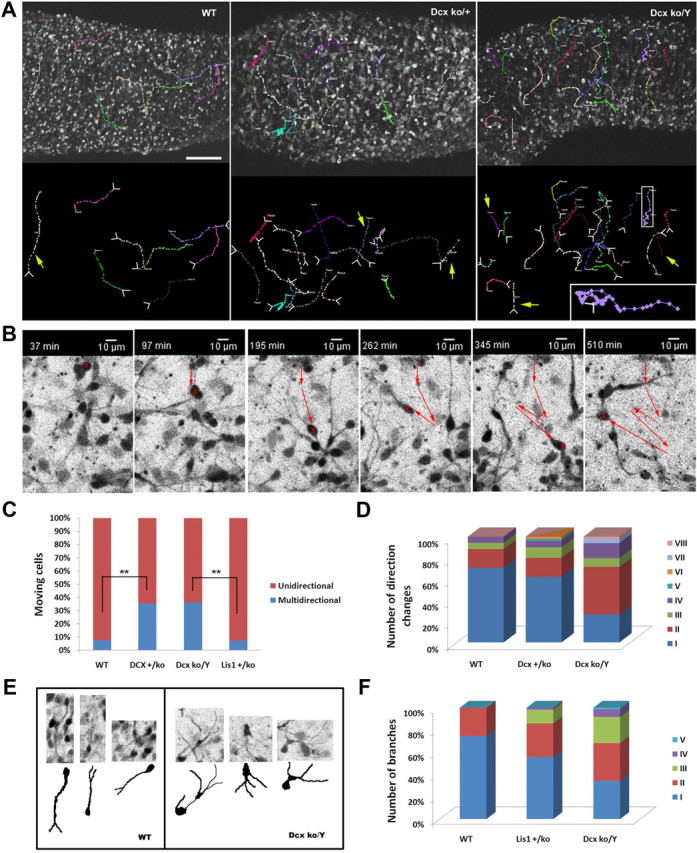
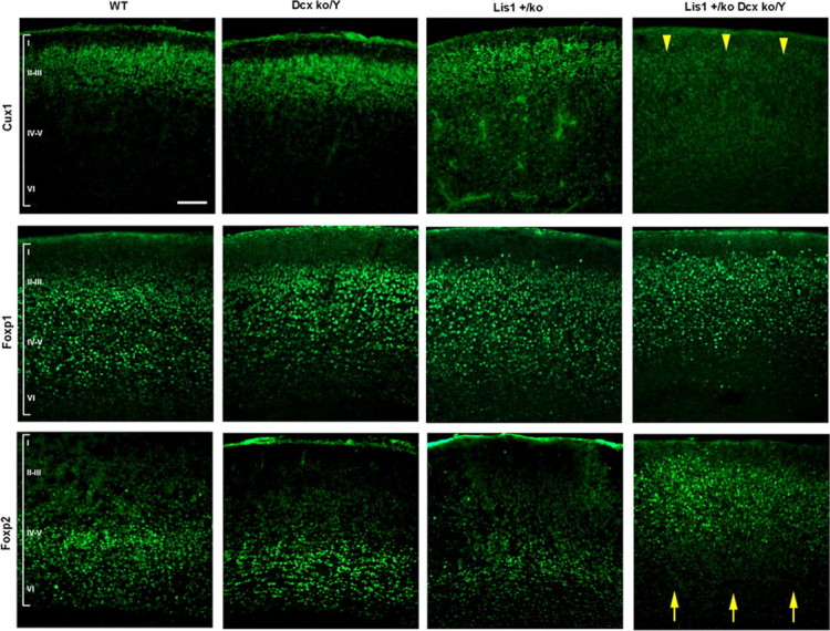
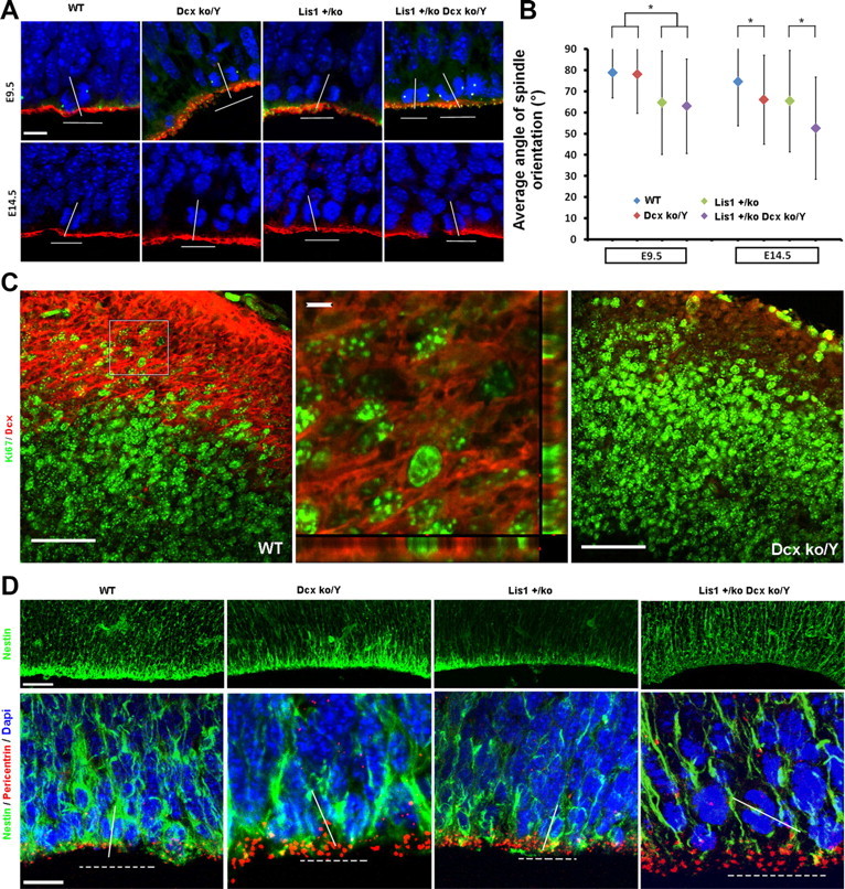
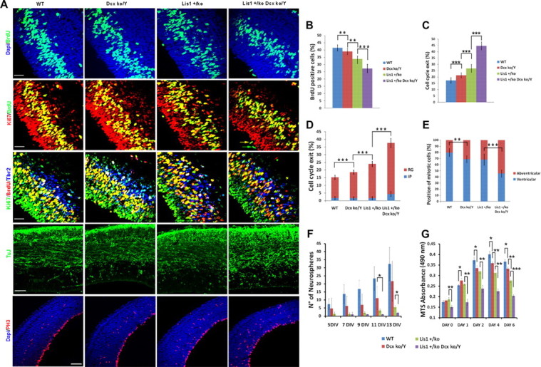
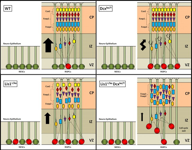
Heterozygous LIS1 mutations and males with loss of the X-linked DCX result in lissencephaly, a neuronal migration defect. LIS1 regulates nuclear translocation and mitotic division of neural progenitor cells, while the role of DCX in cortical development remains poorly understood. Here, we uncovered novel neuronal migration and proliferation defects in the Dcx mutant embryonic brains. Although cortical organization was fairly well preserved, Dcx(ko/Y) neurons displayed defective migration velocities similar to Lis1(+/ko) neurons when characterized by time-lapse video-microscopy of embryonic cortical slices. Dcx(ko/Y) migrating neurons displayed novel multidirectional movements with abnormal morphology and increased branching. Surprisingly, Dcx(ko/Y) radial glial cells displayed spindle orientation abnormalities similar to Lis1(+/ko) cells that in turn lead to moderate proliferation defects both in vivo and in vitro. We found functional genetic interaction of the two genes, with the combined effects of Lis1 haploinsufficiency and Dcx knock-out leading to more severe neuronal migration and proliferation phenotypes in the Lis1(+/ko);Dcx(ko/Y) male double mutant compared with the single mutants, resulting in cortical disorganization and depletion of the progenitor pool. Thus, we provide definitive evidence for a critical role for Dcx in neuronal migration and neurogenesis, as well as for the in vivo genetic interaction of the two genes most commonly involved in human neuronal migration defects.


Figures

Figure 1.
Figure 1.
Lis1 and Dcx double mutants have severe disorganization of the hippocampus, cerebellum, and reduction of the cortical area during development. Morphological analysis at P21, P0, and E13.5 of WT and mutant brain sections. A, Cresyl violet staining and thy1-YFP transgene expression revealed severe morphological abnormalities in the Lis1+/ko;Dcx+/ko female mutants with enlargement of the ventricles, hippocampal disorganization and loss of other brain areas. Scale bar, 1 mm. B, Cresyl violet (I–IV) and Testis-1 staining (V–VIII) of the hippocampus demonstrated an increased severity of disorganization across the mutants. Scale bar, 500 μm. Cerebellar organization (IX–XII) was also severely compromised in the Lis1+/ko;Dcxko/Y male mutant compared with the other genotypes. Scale bar, 100 μm. C, Nissl staining of the hippocampus at P0 confirmed the severe developmental disorganization of the Lis1+/ko;Dcxko/Y male double mutant. Scale bar, 200 μm. Cresyl violet staining of E13.5 brain sections revealed a reduction in the cortical area in the Lis1+/ko;Dcxko/Y male double mutant. Scale bar is 100 μm. The confidence interval indicates the SD. Two-tailed t test distribution was used to calculate p values.
Figure 2.
Figure 2.
Neuronal migration is slower in Dcxko/Y and severely impaired in Lis1+/ko;Dcxko/Y double mutant. Analysis of neuronal migration using organotypic slice cultures. A, Single representative images from time lapse videomicroscopy (supplemental Movies 1–4, available at www.jneurosci.org as supplemental material) demonstrate the gradual decrease of moving cells across the genotypes. The top panel shows the starting position of moving cells with the direction of movement (outlines with arrow). Nonmoving cells (blue outline) remain at approximately the same position until the end of the movie. The bottom panel shows the end position of moving cells and the trajectory they followed (dotted lines). B–D, Quantification of neuronal migration across the genotypes. B, The percentage of moving and stationary cells within three arbitrary areas from each time-lapse video-microscopy movie at the level of the IZ is shown. Cells with an appreciable movement from the starting position were considered to be migrating and were included in the tracking analysis. C, Each movie was calibrated and analyzed to determine the velocity of each single neuron. The average velocity values displayed are independent from the movement direction (for direction-dependent values, see supplemental Fig. 1, available at www.jneurosci.org as supplemental material). D, Velocity values were corrected by combining data from moving and nonmoving cells. Error bars indicate SD; *p < 0.05, ***p < 0.0001 by two-tailed t test.
Figure 3.
Figure 3.
Neuronal migration of Dcx knock-out cortical neurons is multidirectional. Neural precursor cells from Dcx mutant brains displayed a multidirectional phenotype with increased branching and morphological abnormalities. A, Dcx heterozygous and hemizygous mutations cause neurons to migrate with several changes of direction. The starting position and the direction of the movements are indicated by white arrows at the tracking lines. Yellow arrows indicate unidirectional movements. Scale bar, 100 μm. B, Dcx knock-out cell migrated with a zig-zag movement and displayed increased branching. C, Quantification of the number of cells with multidirectional movements. In the Dcx mutants, the proportion of cells with this atypical pattern is at least three times more frequent than WT and Lis1+/ko cells. D, Quantification of the number of direction changes in the multidirectional-migrating cells. The number of changes of direction is higher in the Dcxko/Y mutants than in the Dcx+/ko mutants. E, F, Dcxko/Y migrating neurons displayed increased branching and morphological abnormalities with a swelling protruding or detached from the cell body. **p < 0.001 by two-tailed t test.
Figure 4.
Figure 4.
Cortical organization is severely abnormal in the Lis1+/ko;Dcxko/Y male mutants. Cortical layer-specific immunostaining in each of the genotypes, using Cux1 (layers II–III), Foxp1 (layers III–V) and Foxp2 (layer V–VI) antibodies. Major abnormalities are evident in the Lis1+/ko;Dcxko/Y double mutant with absence of Cux1-positive cells, misplacement of Foxp2-positive cells (yellow arrowheads and arrows). Milder abnormalities are present in the Lis1+/ko mutant with reduction in the number of positive cells. Later born neurons (Cux1 positive) are reduced or absent in the Lis1+/ko;Dcxko/Y double mutant suggesting that there is a neurogenesis defect in addition to defects in neuronal migration. Scale bar, 100 μm.
Figure 5.
Figure 5.
Dcxko/Y and Lis1+/ko;Dcxko/Y male mutants display randomized spindle positioning at the VZ. Analysis of spindle orientation and cellular organization at the ventricular surface. A, Spindle orientation analysis of cells in telophases and immunostaining of aPKC/γ-Tubulin at E9.5 (top; red/green) and aPKC E14.5 (bottom; red). The localization of atypical PKC (red) is normal indicating that apical polarity is preserved. Scale bar, 10 μm. B, Quantification of the average angle of spindle positioning during neuroepithelial and radial glial mitotic divisions. At E9.5 the average spindle orientation is affected only by Lis1 mutation. At E14.5 both Lis1 and Dcx mutants display spindle orientation defects with a more severe randomization in the Lis1+/ko;Dcxko/Y double mutant (*p < 0.05 by two-tailed t test). Error bars indicate SD. C, Double staining with Dcx/Ki67 markers in WT and Dcxko/Y mutant (negative control). DCX expression is highly expressed at the IZ/SVZ in dividing and nondividing cells. In the orthogonal view (second panel, zoom-in), Dcx is localized around the cell. Scale bars: (first and third panel) 50 μm; (second panel) 5 μm. D, Nestin immunostaining suggests that cellular organization is disrupted in the Lis1+/ko;Dcxko/Y male mutant with signal reduction at the VZ and more fragmented fibers. Scale bars, 50 and 10 μm. Pericentrin was used to delineate the apical surface and to define mitotic cells by helping to orient the spindle.
Figure 6.
Figure 6.
In vivo and in vitro proliferation is defective in Lis1+/ko;Dcxko/Y male mutants with depletion of the progenitor pool during development. In vivo and in vitro analysis of proliferation in each of the genotypes at E14.5. A, BrdU immunostaining (top; green) demonstrates a reduction in number of proliferating cells, especially in the Lis1+/ko;Dcxko/Y double mutant. Double with BrdU/Ki-67 antibodies (second row) and triple immunostaining with BrdU/Ki-67/Tbr2 (third row) antibodies was used to measure overall cell cycle exit and cell cycle exit in intermediate progenitor and radial glial cells. The two arrows in the Lis1+/ko;Dcxko/Y double mutant indicate the different fates of the two dividing daughter cells. TuJ immunostaining is stronger in the Lis1+/ko;Dcxko/Y double mutants, consistent with the increase number of neurogenic divisions (fourth panel from top). PH3 immunostaining demonstrates an increasing proportion of mitotic cells dividing away from the ventricular surface in each of the mutants (fifth panel from top). Scale bars: 30 μm for BrdU and cell cycle exit, 50 μm for TuJ staining, 100 μm for PH3 staining. B, Quantification of the BrdU incorporation. C, Quantification of total cell cycle exit using BrdU/Ki-67 double staining. D, Proportion of cells exiting the cell cycle in the intermediate progenitors (IP) and radial glial (RG) cells using BrdU/Ki-67/Tbr2 triple staining. E, Quantification of PH3-positive cells: the proportion of abventricular mitoses increases in each of the mutant genotypes, with the highest proportion present in the Lis1+/ko;Dcxko/Y double mutants. F, Quantification of the number of neurospheres generated per well during 13 d of neurosphere culture. G, Quantification of the MTS absorption during 6 d of neurosphere culture. Error bars indicate SD; *p < 0.05, **p < 0.001, ***p < 0.0001 by two-tailed t test.
Figure 7.
Figure 7.
Summary of neuronal migration and neurogenesis defects found in single Lis1+/ko and Dcxko/Y mutants and double Lis1+/ko;Dcxko/Y mutants. In the WT, neuroepithelial stem cells (NESCs, E9.5 stage) actively proliferate in a stem cell like manner with the spindle orientation mostly perpendicular to the surface. At later stages (E14.5), radial glial progenitors (RGPCs) divide both symmetrically and asymmetrically to generate postmitotic neurons that migrate in an inside-out manner to organize the cortical plate into six layers. Loss of Dcx does not affect spindle positioning during neuroepithelial expansion but does have effects on radial glial progenitor cells, randomizing spindle orientation similar to Lis1 heterozygotes. Although the final cortical organization is preserved in Dcx male mutants, neuronal migration is slower with a multidirectional pattern of migration. Lis1 haploinsufficiency, as previously described (Yingling et al., 2008) results in spindle orientation defects both during neuroepithelial expansion and radial glial mitotic divisions. Cortical organization is mostly preserved with a mild disorganization and a reduction of the number of neurons in the most outer and inner layers of the cortex. In contrast the Lis1+/ko;Dcxko/Y double mutants displayed a more severe randomization of the spindle during radial glial mitotic divisions with abventricular divisions leading to increased cell-cycle exit and depletion of the progenitor pool at the ventricular surface. Neuronal migration and proliferation defects lead to a severe disorganization of the cortex with misplaced neurons and the lack of Cux1-positive cells. In contrast, during neuroepithelial cell divisions the spindle orientation is not significantly different from the Lis1+/ko brains.

Similar articles

Cited by

References

    1. Bai J, Ramos RL, Ackman JB, Thomas AM, Lee RV, LoTurco JJ. RNAi reveals doublecortin is required for radial migration in rat neocortex. Nat Neurosci. 2003;6:1277–1283. - PubMed
    1. Bantubungi K, Blum D, Cuvelier L, Wislet-Gendebien S, Rogister B, Brouillet E, Schiffmann SN. Stem cell factor and mesenchymal and neural stem cell transplantation in a rat model of Huntington's disease. Mol Cell Neurosci. 2008;37:454–470. - PubMed
    1. Brown JP, Couillard-Després S, Cooper-Kuhn CM, Winkler J, Aigner L, Kuhn HG. Transient expression of doublecortin during adult neurogenesis. J Comp Neurol. 2003;467:1–10. - PubMed
    1. Caspi M, Atlas R, Kantor A, Sapir T, Reiner O. Interaction between LIS1 and doublecortin, two lissencephaly gene products. Hum Mol Genet. 2000;9:2205–2213. - PubMed
    1. Corbo JC, Deuel TA, Long JM, LaPorte P, Tsai E, Wynshaw-Boris A, Walsh CA. Doublecortin is required in mice for lamination of the hippocampus but not the neocortex. J Neurosci. 2002;22:7548–7557. - PMC - PubMed

Publication types

MeSH terms