. 2010 Feb 24;30(8):3113-23.
doi: 10.1523/JNEUROSCI.5598-09.2010.

Bral1: its role in diffusion barrier formation and conduction velocity in the CNS

Affiliations

Bral1: its role in diffusion barrier formation and conduction velocity in the CNS

Yoko Bekku et al. J Neurosci. .

Abstract

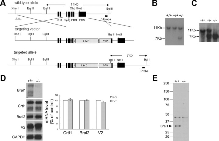
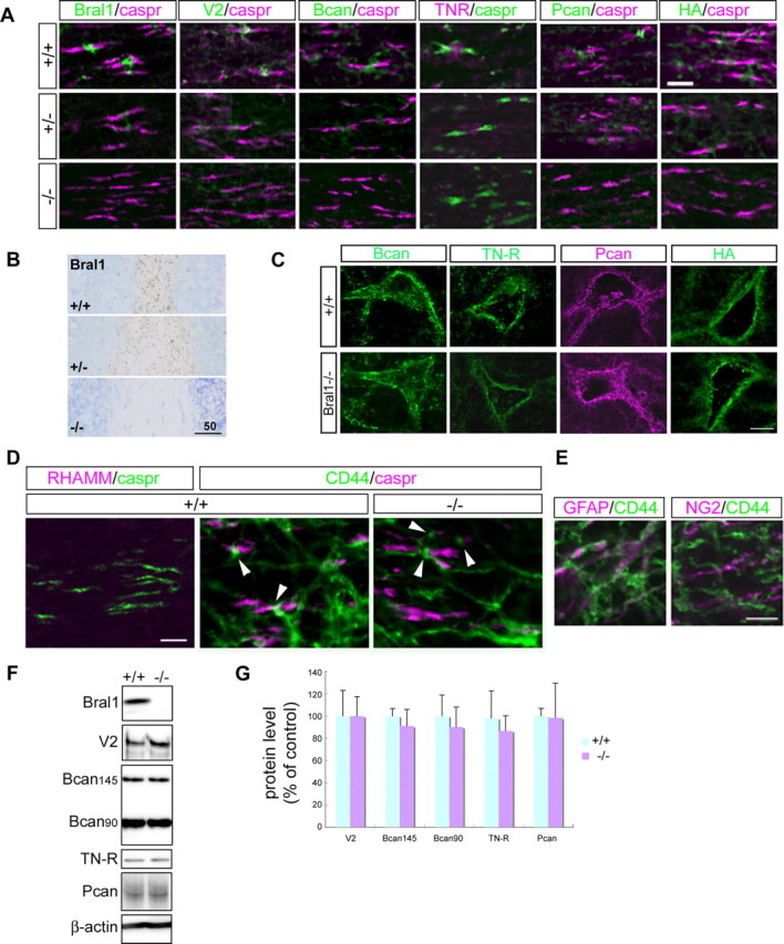
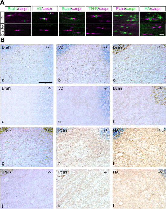
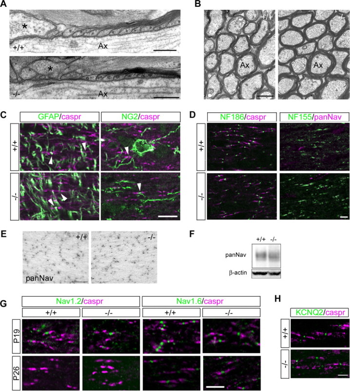
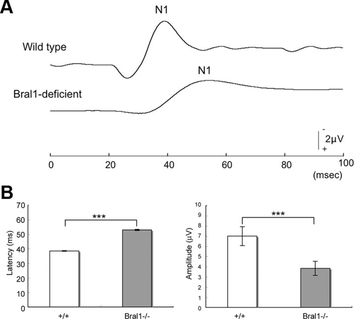
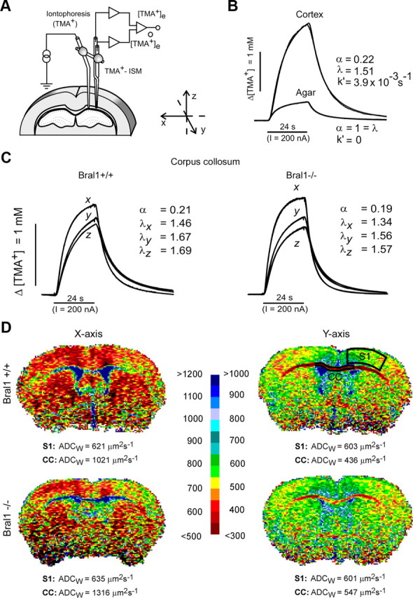
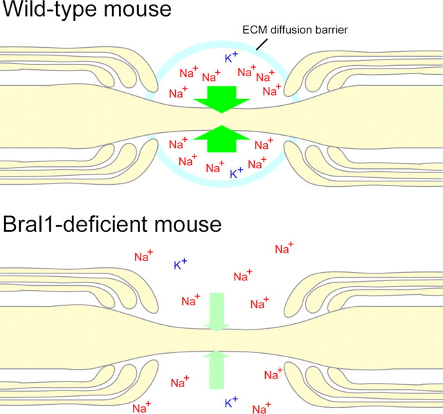
At the nodes of Ranvier, excitable axon membranes are exposed directly to the extracellular fluid. Cations are accumulated and depleted in the local extracellular nodal region during action potential propagation, but the impact of the extranodal micromilieu on signal propagation still remains unclear. Brain-specific hyaluronan-binding link protein, Bral1, colocalizes and forms complexes with negatively charged extracellular matrix (ECM) proteins, such as versican V2 and brevican, at the nodes of Ranvier in the myelinated white matter. The link protein family, including Bral1, appears to be the linchpin of these hyaluronan-bound ECM complexes. Here we report that the hyaluronan-associated ECM no longer shows a nodal pattern and that CNS nerve conduction is markedly decreased in Bral1-deficient mice even though there were no differences between wild-type and mutant mice in the clustering or transition of ion channels at the nodes or in the tissue morphology around the nodes of Ranvier. However, changes in the extracellular space diffusion parameters, measured by the real-time iontophoretic method and diffusion-weighted magnetic resonance imaging (MRI), suggest a reduction in the diffusion hindrances in the white matter of mutant mice. These findings provide a better understanding of the mechanisms underlying the accumulation of cations due to diffusion barriers around the nodes during saltatory conduction, which further implies the importance of the Bral1-based extramilieu for neuronal conductivity.


Figures

Figure 1.
Figure 1.
Targeted disruption of the Bral1 gene. A, Bral1 targeting strategy. The wild-type Bral1 gene (top), the targeting vector (middle), and the disrupted Bral1 gene (bottom) are shown. The expected fragment sizes after BglII digestion and hybridization with a probe are 11 kb for the wild-type allele and 7 kb for the recombinant allele. The asterisk indicates the location of the exon encoding the peptide used as the immunogen for the Bral1 antibody. UT, Untranslated region; Sp, signal peptide; Ig, Ig-fold; PTR, proteoglycan tandem repeat. B, Southern blot analysis of genomic DNA from ES cell clones shows the wild-type (11 kb) and the targeted (7 kb) alleles. C, Southern blot analysis of tail DNA isolated from a mouse homozygous for the wild-type allele (+/+), a heterozygous mouse (+/−), and a homozygous mutant mouse (−/−). D, Northern blot analysis of polyA(+) RNA isolated from the brain of wild-type, heterozygous (+/−), and homozygous mutant (−/−) mice. The blot was sequentially hybridized with probes specific for mouse Bral1, Crtl1, Bral2, versican V2, and Gapdh. In quantification of those transcripts, blots were scanned and densitometric analysis was performed using the Gapdh signal to normalize mRNA levels. Data are presented as a percentage of wild-type levels. Error bars indicate ±SD. E, Western blot analysis of crude extracts (80 μg) from wild-type (+/+) and Bral1-deficient (−/−) mouse brains. Samples were resolved on a 2–15% gradient SDS-PAGE gel and immunoblotted with an antibody directed against Bral1.
Figure 2.
Figure 2.
Bral1 is indispensable for stabilizing extracellular matrices at the nodes of Ranvier in the CNS. A, Immunofluorescent labeling of the optic nerve in wild-type mice (+/+; upper column), heterozygous mice (+/−; middle column), and Bral1-deficient mice (−/−; lower column) using antibodies to Bral1 (green), versican V2 (green), brevican (Bcan/green), TN-R (magenta), phosphacan (Pcan/green), and caspr (magenta or green), or labeling with B-HABP (HA/green). Note that all extracellular matrices involving Bral1 no longer show a nodal pattern in the CNS of Bral1-deficient mice. B, Immunolabeling of the white matter in the cerebellum using an antibody to Bral1. Note that the expression level is decreased even though the expression pattern is not affected in the heterozygous mouse. C, Immunolabeling of perineuronal nets (PNN) with antibodies against brevican (Bcan/green), TN-R (green), and phosphacan (Pcan/magenta) or labeling with B-HABP (HA/green). The expression of these molecules in PNN was not affected in Bral1-deficient mice. D, Immunofluorescent labeling of the optic nerves in wild-type mice (+/+) and Bral1-deficient mice (−/−) using antibodies directed against the hyaluronan receptors RHAMM (magenta) and CD44 (green). Note that CD44-positive processes (arrowhead) project to some nodal gaps and that the expression is not affected in Bral1-deficient mice. E, Glial cell type that expresses CD44 in the optic nerve. Note that the GFAP-positive astrocyte (magenta) expresses CD44 (green). F, Western blot analysis of the extracellular matrix in wild-type and Bral1-deficient mice. Crude extracts (15 μg) from wild-type (n = 5) and Bral1-deficient (n = 6) mouse optic nerves were resolved on a 2–15% gradient SDS-PAGE gel and immunoblotted with antibodies to Bral1, versican V2, brevican, and TN-R. G, To quantify the protein levels, densitometric analysis was performed. Data are presented as a percentage of wild-type levels. Error bars indicate ±SD. Scale bars: A, 10 μm; B, 50 μm; C, 20 μm; D, E, 5 μm.
Figure 3.
Figure 3.
Complete absence of the nodal ECM in the CNS of Bral1-deficient mice. A, Immunofluorescent labeling of the facial nerve tract in wild-type mice (upper column) and Bral1-deficient mice (lower column) using antibodies to Bral1 (green), versican V2 (green), brevican (Bcan/green), TN-R (green), phosphacan (Pcan/magenta), and caspr (magenta or green) or labeling with B-HABP (HA/green). B, Immunohistochemical staining of cerebellar sections from wild-type mice (+/+) and Bral1-deficient mice (−/−) using antibodies against Bral1 (a, d), versican V2 (b, e), brevican (c, f), TN-R (g, j), and phosphacan (h, k) or B-HABP-labeled hyaluronan (i, l). Scale bars: A, 10 μm; B, 100 μm.
Figure 4.
Figure 4.
The nodal component structure was not affected in Bral1-deficient mice. A, Ultrastructural analysis of the optic nerves. Longitudinal sections through the optic nerves of 6-month-old wild-type (+/+) and Bral1-deficient (−/−) mice. Note the normal ultrastructure of the paranodal regions of the myelin sheaths and the presence of perinodal astrocyte processes (some marked with asterisk) extending into the nodal regions of the axons. B, Cross sections of wild-type (+/+) and Bral1-deficient (−/−) mice. There are no significant differences in the number of myelinated axons or the ultrastructure of myelin between the two genotypes. Ax, Axon. C, Immunofluorescent analysis of the perinodal glial processes in the optic nerve of wild-type (+/+) and Bral1-deficient (−/−) mice. There are no differences in the projection to nodal gaps of either GFAP-positive astrocytes (arrowhead in left column) or NG2 glia (arrowhead in right column) between wild-type and knock-out animals. D, The distribution of the nodal proteins NF 186 and NF 155 is also not affected in Bral1-deficient mice. E, Immunohistochemical evaluation of sodium channel cluster distribution in the optic nerves of wild-type and Bral1-deficient mice. Representative expression pattern of sodium channel clusters in wild-type (+/+) and Bral1-deficient (−/−) mice. F, Representative data of Western blot, which was performed using crude extracts (15 μg) from the optic nerves of wild-type and Bral1-deficient mice. G, Sodium channel subtype transition occurs normally in Bral1-deficient mice. H, The clustering of KCNQ2 was also not affected in Bral1-deficient mice. Scale bars: A, 300 nm; B, 750 nm; C, E, 10 μm; D, G, H, 5 μm.
Figure 5.
Figure 5.
fVEPs were affected in Bral1-deficient mice. A, Representative waveforms of VEP from a wild-type mouse (upper waveform) and a Bral1-deficient mouse (lower waveform). Note that the N1 peak of the Bral1-deficient mouse was lower and prolonged. B, Summary of VEP from wild-type (n = 5) and Bral1-deficient (n = 6) mice. Note that the latency was extremely increased and the amplitude was significantly decreased in Bral1-deficient mice. Significance value was calculated using Student's t test; ***p < 0.001. Error bars indicate ±SD.
Figure 6.
Figure 6.
The diffusion properties of the ECS in the corpus callosum. A, Experimental arrangement. Tetramethylammonium ions (TMA+) were iontophoresed into the tissue by an iontophoretic micropipette, and their concentration was measured at a known distance by a TMA+-selective microelectrode. The micropipette and microelectrode were glued together to stabilize the intertip distance. B, Typical diffusion curves evoked by TMA+ iontophoresis in agar and in the cortex. The theoretical diffusion curves generated by a nonlinear curve-fitting simplex algorithm are superimposed on the actual diffusion curves recorded in the tissue or agar. Before tissue measurements, several diffusion curves were recorded in agar, where by definition α = 1 = λ and k′ = 0, thus enabling the transport number of the electrode array to be determined. In the tissue, the resulting increase in concentration was much larger than that in agar due to the restricted volume fraction and increased tortuosity in the brain. C, Examples of the TMA+ diffusion curves recorded in the corpus callosum of Bral1-positive and -negative mice. In contrast to diffusion in the cortex, which is isotropic, meaning that the values of the ECS diffusion parameters are the same along all three orthogonal axes, in the anisotropic corpus callosum, there is preferential diffusion along the myelinated fibers (x-axis). The different diffusion curves resulting from the unequal tortuosity values measured along the three orthogonal axes indicate anisotropic diffusion in both wild-type and knock-out mice; however, tortuosity λ in mutant mice was decreased along all the main axes (values λx, λy, and λz). D, Typical ADCW maps of wild-type and Bral1-deficient mice along the mediolateral (x) and rostrocaudal (y) axes. The mean value of ADCW, given below each map, was calculated in the outlined regions of interest. The calculated ADCW values were significantly higher in Bral1 knock-out mice than in controls in the CC but not in the primary somatosensory cortex (S1). The scale shows the relation between the intervals of ADCW values and the colors used for visualization; the values shown on the left are valid for the x-axis, those on the right for the y-axis.
Figure 7.
Figure 7.
A hypothetical model for the nodal diffusion barrier in the CNS. Hyaluronan-bound gel-like matrices would maintain the microenvironment and function as an ion diffusion barrier around the perinodal ECS in wild-type mice. The ion diffusion barrier would have a dramatic effect on the local accumulation of Na+ and K+ ions in the extracellular nodal region. On the other hand, the destruction of the nodal ECM caused by Bral1 deficiency allows ions to diffuse at the perinodal ECS region.

References

    1. Asher RA, Scheibe RJ, Keiser HD, Bignami A. On the existence of a cartilage-like proteoglycan and link proteins in the central nervous system. Glia. 1995;13:294–308. - PubMed
    1. Aspberg A, Miura R, Bourdoulous S, Shimonaka M, Heinegârd D, Schachner M, Ruoslahti E, Yamaguchi Y. The C-type lectin domains of lecticans, a family of aggregating chondroitin sulfate proteoglycans, bind tenascin-R by protein-protein interactions independent of carbohydrate moiety. Proc Natl Acad Sci U S A. 1997;94:10116–10121. - PMC - PubMed
    1. Bekku Y, Su WD, Hirakawa S, Fässler R, Ohtsuka A, Kang JS, Sanders J, Murakami T, Ninomiya Y, Oohashi T. Molecular cloning of Bral2, a novel brain-specific link protein, and immunohistochemical colocalization with brevican in perineuronal nets. Mol Cell Neurosci. 2003;24:148–159. - PubMed
    1. Bekku Y, Rauch U, Ninomiya Y, Oohashi T. Brevican distinctively assembles extracellular components at the large diameter nodes of Ranvier in the CNS. J Neurochem. 2009;108:1266–1276. - PubMed
    1. Boiko T, Rasband MN, Levinson SR, Caldwell JH, Mandel G, Trimmer JS, Matthews G. Compact myelin dictates the differential targeting of two sodium channel isoforms in the same axon. Neuron. 2001;30:91–104. - PubMed

Publication types

MeSH terms