. 2010 Feb 24;30(8):3124-32.
doi: 10.1523/JNEUROSCI.5848-09.2010.

Neurokinin B and dynorphin A in kisspeptin neurons of the arcuate nucleus participate in generation of periodic oscillation of neural activity driving pulsatile gonadotropin-releasing hormone secretion in the goat

Affiliations

Neurokinin B and dynorphin A in kisspeptin neurons of the arcuate nucleus participate in generation of periodic oscillation of neural activity driving pulsatile gonadotropin-releasing hormone secretion in the goat

Yoshihiro Wakabayashi et al. J Neurosci. .

Abstract

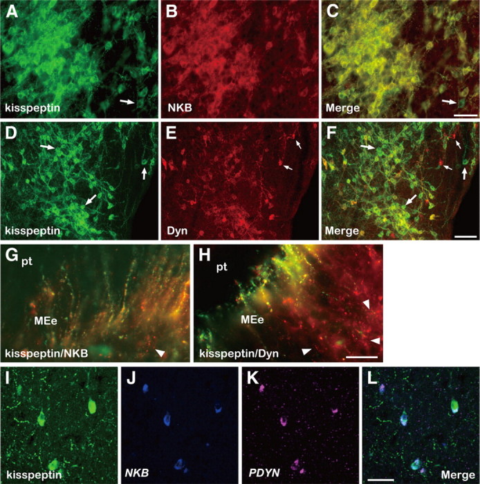
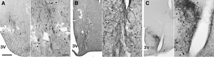
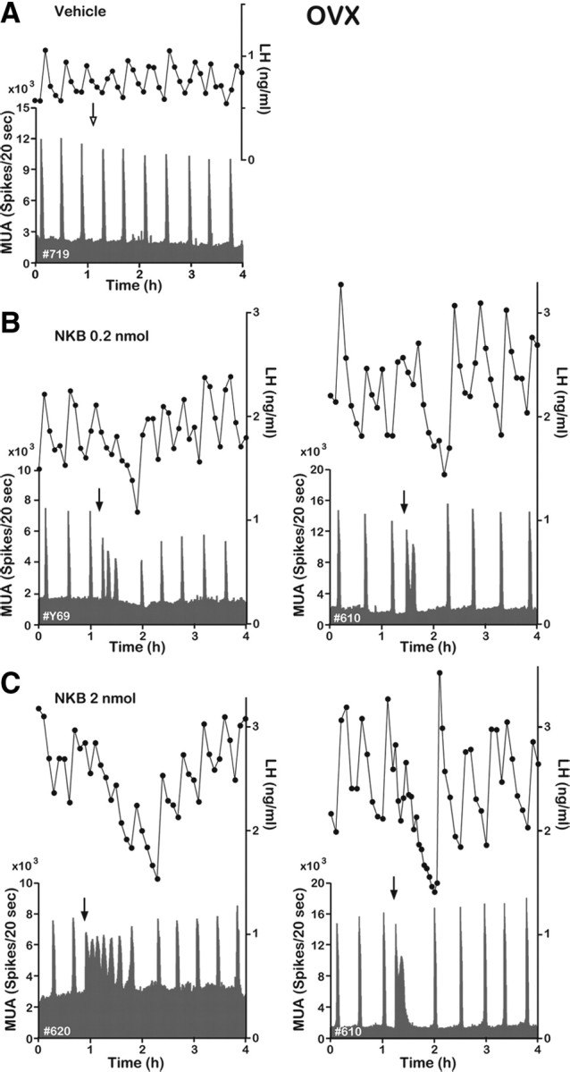
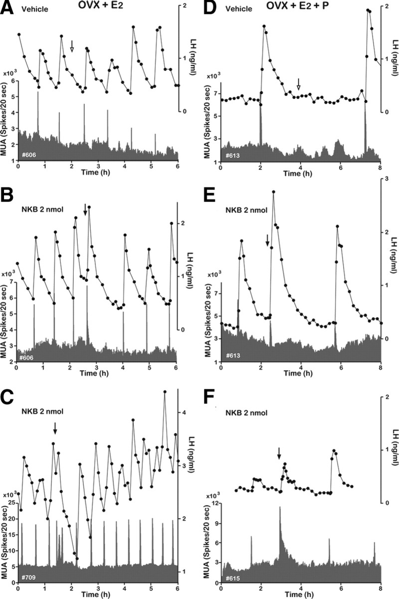
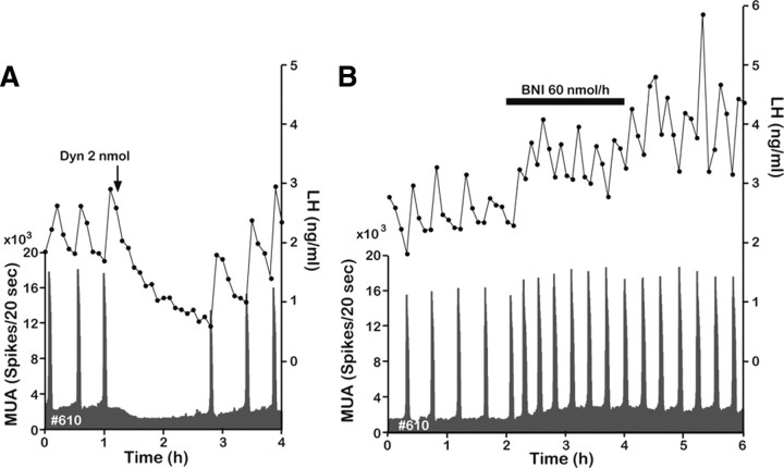
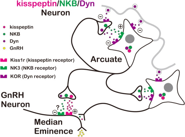
Gonadotropin-releasing hormone (GnRH) neurons in the basal forebrain are the final common pathway through which the brain regulates reproduction. GnRH secretion occurs in a pulsatile manner, and indirect evidence suggests the kisspeptin neurons in the arcuate nucleus (ARC) serve as the central pacemaker that drives pulsatile GnRH secretion. The purpose of this study was to investigate the possible coexpression of kisspeptin, neurokinin B (NKB), and dynorphin A (Dyn) in neurons of the ARC of the goat and evaluate their potential roles in generating GnRH pulses. Using double and triple labeling, we confirmed that all three neuropeptides are coexpressed in the same population of neurons. Using electrophysiological techniques to record multiple-unit activity (MUA) in the medial basal hypothalamus, we found that bursts of MUA occurred at regular intervals in ovariectomized animals and that these repetitive bursts (volleys) were invariably associated with discrete pulses of luteinizing hormone (LH) (and by inference GnRH). Moreover, the frequency of MUA volleys was reduced by gonadal steroids, suggesting that the volleys reflect the rhythmic discharge of steroid-sensitive neurons that regulate GnRH secretion. Finally, we observed that central administration of Dyn-inhibit MUA volleys and pulsatile LH secretion, whereas NKB induced MUA volleys. These observations are consistent with the hypothesis that kisspeptin neurons in the ARC drive pulsatile GnRH and LH secretion, and suggest that NKB and Dyn expressed in those neurons are involved in the process of generating the rhythmic discharge of kisspeptin.


Figures

Figure 1.
Figure 1.
Colocalization of kisspeptin, NKB, and Dyn in the caudal ARC and ME of the goat. Photomicrographs of sections of the ARC stained by immunocytochemistry for kisspeptin (A) and NKB (B), or kisspeptin (D) and Dyn (E), are shown. C and F are computer-aid merged images of A and B, or D and E, respectively. An arrow in A or C indicates a cell body containing immunoreactivity (ir) for kisspeptin but not NKB-ir. The large arrows in D or F, or small arrows in E or F, show some cell bodies containing exclusively kisspeptin-ir or Dyn-ir, respectively. Note that numerous kisspeptin/NKB- or kisspeptin/Dyn-positive fibers surround immunopositive cell bodies. G and H are merged images of sections of the ME double stained for kisspeptin/NKB or kisspeptin/Dyn, respectively. The arrowhead in G indicates a fiber with only NKB-ir. The arrowheads in H show fibers with exclusively Dyn-ir. I–K show kisspeptin-ir and positive signals for NKB and PDYN in triple-label histochemistry, respectively. L is a merged image of I–K. MEe, External layer of median eminence; pt, pars tuberalis. Scale bars: A–C, I–L, 50 μm; D–F, 100 μm; G, H, 25 μm.
Figure 2.
Figure 2.
Photomicrographs showing the placement of MUA recording electrodes in three goats [no. Y69 (A), no. 709 (B), and no. 713 (C)] from which MUA was successfully recorded. Sections were immunostained for kisspeptin. A pair of brackets indicates the area where a trace of a bundle of electrodes is observed, and a magnification of the indicated area is shown on the right side of each panel. Some kisspeptin-immunopositive cell bodies are shown by arrowheads. Note that because it was not possible to perfuse goats Y69 and 709, immunostaining of their sections was poor. 3V, Third ventricle. Scale bars: left panels, 500 μm; right panels, 100 μm.
Figure 3.
Figure 3.
Effect of NKB on the MUA and plasma LH in the OVX goat. Representative profiles of the MUA and plasma LH concentrations in OVX animals that received an intracerebroventricular injection of vehicle (A) or 0.2 (B) or 2 nmol (C) of NKB are shown. The arrow indicates timing of injection.
Figure 4.
Figure 4.
Effect of NKB on MUA and plasma LH in OVX-plus-E2- and OVX-plus-E2-plus-P-treated goats. Representative profiles of MUA and plasma LH concentrations in OVX-plus-E2 (A–C) and OVX-plus-E2-plus-P (D–F) goats that received an intracerebroventricular injection of vehicle (A, D) or 2 nmol (B, C, E, F) of NKB are shown. The arrow indicates timing of injection.
Figure 5.
Figure 5.
Effects of Dyn and nor-BNI on the MUA and plasma LH in the OVX goat. A, Representative profiles of MUA and plasma LH concentrations in one animal that received an intracerebroventricular injection of 2 nmol of Dyn are shown. The arrow indicates timing of injection. B, Representative profiles of MUA and plasma LH concentrations in one animal that received an intracerebroventricular infusion of nor-BNI at a rate of 60 nmol · 600 μl−1 · h−1 for 2 h are shown. The bar indicates the period of infusion.
Figure 6.
Figure 6.
Schematic representation of the role of ARC kisspeptin/NKB/Dyn neurons in the generation of the pulsatile GnRH release. According to this model, kisspeptin/NKB/Dyn neurons in the ARC form a neural circuit by their collaterals and dendrites. Within the neural circuit, NKB/NK3 signaling plays the role of accelerator, whereas Dyn/KOR signaling serves as a brake on activation of kisspeptin/NKB/Dyn neurons. Through the reciprocal actions of NKB/NK3 and Dyn/KOR signaling, rhythmic oscillation of neural activity is generated in kisspeptin/NKB/Dyn neurons, which in turn induces pulsatile kisspeptin release at the ME and hence pulsatile GnRH release into the portal circulation. Thus, ARC kisspeptin/NKB/Dyn neurons would act as the GnRH pulse generator through the coordinated interaction between three peptides. See text for details.

References

    1. Belchetz PE, Plant TM, Nakai Y, Keogh EJ, Knobil E. Hypophysial responses to continuous and intermittent delivery of hypopthalamic gonadotropin-releasing hormone. Science. 1978;202:631–633. - PubMed
    1. Brown CH, Ludwig M, Leng G. κ-Opioid regulation of neuronal activity in the rat supraoptic nucleus in vivo. J Neurosci. 1998;18:9480–9488. - PMC - PubMed
    1. Burke MC, Letts PA, Krajewski SJ, Rance NE. Coexpression of dynorphin and neurokinin B immunoreactivity in the rat hypothalamus: morphologic evidence of interrelated function within the arcuate nucleus. J Comp Neurol. 2006;498:712–726. - PubMed
    1. Dellovade TL, Merchenthaler I. Estrogen regulation of neurokinin B gene expression in the mouse arcuate nucleus is mediated by estrogen receptor alpha. Endocrinology. 2004;145:736–742. - PubMed
    1. Foradori CD, Coolen LM, Fitzgerald ME, Skinner DC, Goodman RL, Lehman MN. Colocalization of progesterone receptors in parvicellular dynorphin neurons of the ovine preoptic area and hypothalamus. Endocrinology. 2002;143:4366–4374. - PubMed

Publication types

MeSH terms