. 2010 Jun;82(6):1227-36.
doi: 10.1095/biolreprod.109.079202. Epub 2010 Feb 24.

EPAS1 Is Required for Spermatogenesis in the Postnatal Mouse Testis

Affiliations

EPAS1 Is Required for Spermatogenesis in the Postnatal Mouse Testis

Michaela Gruber et al. Biol Reprod. 2010 Jun.

Abstract

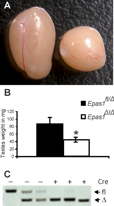
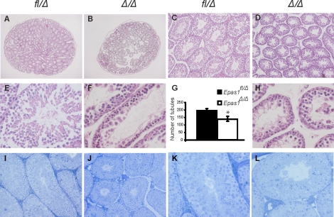
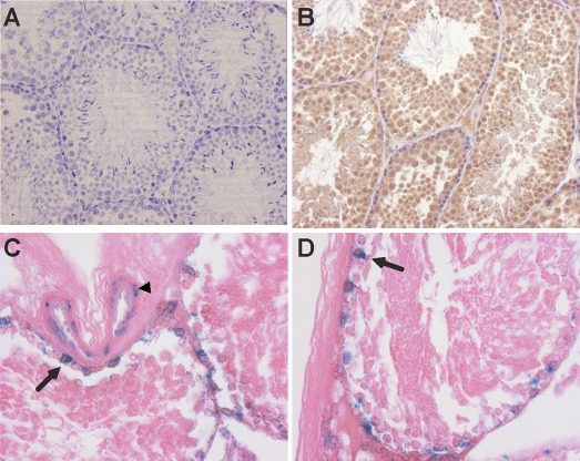
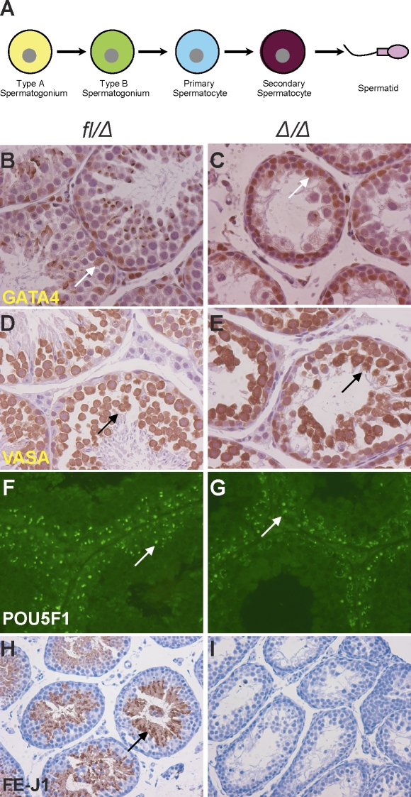
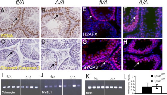
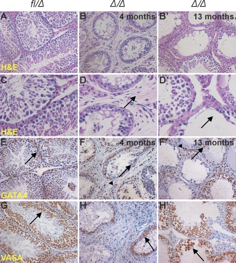
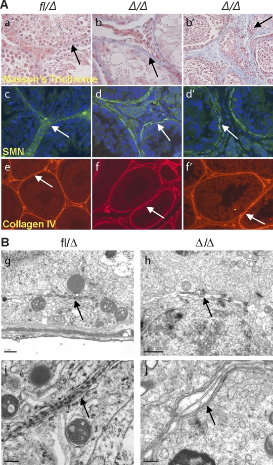
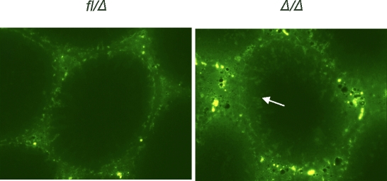
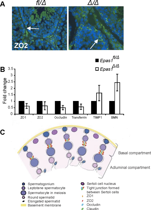
Spermatogenesis, a process involving the differentiation of spermatogonial stem cells into mature spermatozoa, takes place throughout masculine life. A complex system in the testis, including endocrine signaling, physical interactions between germ and somatic cells, spermatocyte meiosis, and timely release of spermatozoa, controls this cycle. We demonstrate herein that decreased O(2) levels and Epas1 activation are critical components of spermatogenesis. Postnatal Epas1 ablation leads to male infertility, with reduced testis size and weight. While immature spermatogonia and spermatocytes are present in Epas1(Delta/Delta) testes, spermatid and spermatozoan numbers are dramatically reduced. This is not due to germ cell-intrinsic defects. Rather, Epas(Delta/Delta) Sertoli cells exhibit decreased ability to form tight junctions, thereby disrupting the blood-testis barrier necessary for proper spermatogenesis. Reduced numbers of tight junction complexes are due to decreased expression of multiple genes encoding tight junction proteins, including TJP1 (ZO1), TJP2 (ZO2), and occludin. Furthermore, Epas1(Delta/Delta) testes exhibit disrupted basement membranes surrounding the seminiferous tubules, causing the premature release of incompletely differentiated germ cells. We conclude that low O(2) levels in the male gonad regulate germ cell homeostasis in this organ via EPAS1.


Figures

FIG. 1.
FIG. 1.
Epas1Δ/Δ testes are reduced in size and weight. A) The control testis (Epas1fl/Δ) on the left was approximately twice the size of the Epas1Δ/Δ on the right at age 8 wk. Well-defined vasculature was visible at the testis surface of either genotype. Original magnification ×3. B) Control testes weighed approximately 80 mg in a 30-g mouse. In contrast, Epas1Δ/Δ testes weighed 40 mg each in males displaying the same body weight (n = 5, *P < 0.05). C) Efficient Epas1 deletion was confirmed by PCR for the floxed and deleted allele of DNA isolated from control and Epas1Δ/Δ testes as previously described [25].
FIG. 2.
FIG. 2.
Loss of Epas1 resulted in fewer and smaller seminiferous tubules. A and B) Cross-sections through the center of control and Epas1Δ/Δ testes are displayed. The average number of tubules was enumerated and found to be reduced by 25% in Epas1Δ/Δ testes (n = 5, *P < 0.05) (G). C and D) Epas1Δ/Δ seminiferous tubules have a smaller diameter compared with control testes. E, F, and H) Higher magnification of H&E-stained tissue shows the presence of spermatogonia, spermatocytes, spermatids, and Sertoli cells in control samples but revealed decreased numbers of spermatids in Epas1Δ/Δ testes. Cross-sections of control (I and K) and Epas1Δ/Δ (J and L) testes after fixation by whole-body perfusion and staining with toludine blue also demonstrated the smaller size of mutant tubules. Original magnification ×20 (A and B), ×100 (C and D), × 200 (I and J), and ×400 (E, F, H, K, and L).
FIG. 3.
FIG. 3.
The testis is a naturally “hypoxic” tissue as determined by pimonidazole staining. Protein thiol adducts recognized by immunohistochemical staining occur in cells experiencing less than 1.5% O2. B) Almost all germ cells exhibited positive staining, confirming low O2 levels, whereas no staining was observed in testis of animals not injected with pimonidazole (A). C and D) β-Galactosidase activity detected in frozen sections from heterozygous Epas1:LacZ knockin testes demonstrates Epas1 expression in the nuclei of Sertoli cells (arrows) and blood vessel ECs (arrowhead) but not in germ cells. Original magnification ×200 (A and B) and ×400 (C and D).
FIG. 4.
FIG. 4.
EPAS1Δ/Δ testes exhibit decreased production of spermatids. A) Schematic diagram depicting discrete spermatogenesis. Mitotic divisions of type A and B spermatogonia lead to formation of spermatocytes, followed by meiotic divisions to form spermatids. B) GATA4 expression revealed the localization of Sertoli cells attached to the basement membrane and interspersed with germ cells in controls (arrow). C) Similar Sertoli cell numbers and position were observed in Epas1Δ/Δ animals (n = 3–5) (arrow). D and E) VASA, a cytoplasmic protein expressed in germ cells, confirmed the presence of several layers of germ cells in Epas1fl/Δ and Epas1Δ/Δ testes (arrows). E) Of note, Epas1Δ/Δ germ cells displayed a disorganized architecture relative to controls. To better assess germ cell types present after Epas1 ablation, sections were stained for POU5F1 protein expressed in spermatogonia. F and G) Both control and Epas1Δ/Δ mice harbored equal numbers of POU5F1+ spermatogonia (arrows). H and I) Haploid spermatids were detected by FE-J1 (a component of the acrosomal complex), staining in control testes (arrow). However, Epas1Δ/Δ testes were largely devoid of FE-J1-expressing spermatids. Original magnification ×200 (BE), ×400 (F and G), and ×100 (H and I).
FIG. 5.
FIG. 5.
Proliferation, apoptosis, meiosis, and testosterone levels are not altered in Epas1Δ/Δ testes. A and B) Proliferation was not reduced in response to Epas1 deficiency as assessed by IHC for PCNA1 (n = 4) (arrows). C and D) Cleaved caspase-3 was used as a means to visualize apoptotic cells. Epas1Δ/Δ testes did not show increased numbers of dead cells compared with the control testes (arrows). γ-H2AX staining detects double-strand breaks during crossover and remains present in the XY body. E and F) Epas1fl/Δ and Epas1Δ/Δ testes displayed a similar staining pattern (arrows). SCP-3 expression connecting the two sister chromatids was used as a second marker for meiosis. G and H) Testes of both genotypes contained multiple layers of spermatocytes undergoing meiosis (n = 4) (arrows). IK) The RT-PCR analysis for calmegin (Clgn), Mybl1, and Gpd (used as a loading control) revealed normal expression of these mRNAs. L) Testosterone levels were not changed in Epas1-deficient males. Original magnification ×400 (AH).
FIG. 6.
FIG. 6.
The testicular phenotype progresses with age in Epas1Δ/Δ mice. A) Histological analyses of 4-mo-old and 13-mo-old testes obtained from multiple Epas1-deficient males compared with control Epas1fl/Δ testes (A and C). The H&E staining depicted a phenotype worse than that observed at age 2 mo. Epas1Δ/Δ seminiferous tubules had lost increased numbers of germ cells, and the interstitial space was fibrotic (B, B′, D, and D′ [arrows]). GATA4 and VASA expression was used to identify Sertoli cells (EF′ [arrows]) and germ cells (GH′ [arrows]); both were still present. GATA4 also detected Leydig cells in the interstitial space (EF′ [arrowheads]). Original magnification ×200 (AB′ and EH′) and ×400 (CD′).
FIG. 7.
FIG. 7.
EpasΔ/Δ testes exhibit defective basement membrane and myoid cell layers. A) Masson trichrome staining confirmed the fibroblastic character in the interstitial space of Epas1Δ/Δ mice (a, arrow) (original magnification ×400). Epas1Δ/Δ testes aged 4 mo (b) and 13 mo (b′) are shown. Myoid cells were detected by SMA IF (c); control seminiferous tubules were surrounded by a single layer of continuous myoid cells, promoting a tight basement membrane (arrow) (original magnification ×400). However, Epas1Δ/Δ testes displayed thickening and disruption of the basement membrane (d and d′, arrows) (original magnification ×400); the fibroblast-like cells in the interstitial space did not express SMN. Collagen type IV staining delineated irregularity and thickening of the basement membrane of mutant tubules (ef′, arrows) (original magnification ×200). B) Electron microscopy displayed tight junctions and an impermeable seal between the membranes of two Sertoli cells in control testes (g and i, arrows). Epas1Δ/Δ testes contained shortened tight junctions (arrow in h) and did not form a proper barrier or seal (arrow in j). Furthermore, cells within Epas1Δ/Δ seminiferous tubules were surrounded by multiple membranes (arrow in j). Bar = 1 μm (g, h) and 0.5 μm (i, j).
FIG. 8.
FIG. 8.
Integrity of the BTB is impaired in Epas1Δ/Δ testes. The functional integrity assay displayed increased diffusion of FITC into the seminiferous tubules of Epas1Δ/Δ testes (arrow). In direct contrast, FITC staining is confined to the basal region of the seminiferous epithelium of control testes. Original magnification ×400.
FIG. 9.
FIG. 9.
Tight junction proteins are affected in Epas1Δ/Δ testes. A) ZO2 protein was still detected by IF in both control and Epas1Δ/Δ testes (arrows, original magnification ×400). B) The mRNAs encoding ZO1, ZO2, occludin, and transferrin were somewhat decreased in Epas1Δ/Δ testes. In contrast, the protease inhibitor TIMP1 and SMN (reflecting the myoid cell phenotype) were slightly increased in Epas1Δ/Δ mice (n = 4). Changes in relative abundance of these transcripts were not statistically significantly; however, all four mutant animals clearly displayed a trend toward decreased expression of each gene. C) Schematic drawing showing the different cell types, junctions, and compartments present in the seminiferous tubules. Sertoli cell nuclei (dark pink) are located close to the basement membrane (yellow), but their cytoplasm (light pink) reaches down to the lumen and connects with other Sertoli cells and germ cells. Tight junctions are formed between two Sertoli cells via ZO1, ZO2, occludin, and claudin interaction and divide the seminiferous tubule into a basal and adluminal compartment. Germ cells move toward the lumen as they mature. Spermatogonia (dark blue) are attached to the basement membrane. Once germ cells enter meiosis, they have passed the BTB and are now separated from blood and lymph. Mature spermatozoa are released into the lumen (light blue).

Similar articles

Cited by

References

    1. Vergouwen RP, Huiskamp R, Bas RJ, Roepers-Gajadien HL, Davids JA, de Rooij DG.Postnatal development of testicular cell populations in mice. J Reprod Fertil 1993; 99: 479–485. - PubMed
    1. Dym M, Fawcett DW.The blood-testis barrier in the rat and the physiological compartmentation of the seminiferous epithelium. Biol Reprod 1970; 3: 308–326. - PubMed
    1. Griswold MD.Interactions between germ cells and Sertoli cells in the testis. Biol Reprod 1995; 52: 211–216. - PubMed
    1. Russell LD, Peterson RN.Sertoli cell junctions: morphological and functional correlates. Int Rev Cytol 1985; 94: 177–211. - PubMed
    1. Wenger RH, Katschinski DM.The hypoxic testis and post-meiotic expression of PAS domain proteins. Semin Cell Dev Biol 2005; 16: 547–553. - PubMed

Publication types

MeSH terms