. 2010 May;298(5):F1205-13.
doi: 10.1152/ajprenal.00708.2009. Epub 2010 Feb 24.

Acute regulation of renal Na+/H+ exchanger NHE3 by dopamine: role of protein phosphatase 2A

Affiliations

Acute regulation of renal Na+/H+ exchanger NHE3 by dopamine: role of protein phosphatase 2A

I Alexandru Bobulescu et al. Am J Physiol Renal Physiol. 2010 May.

Abstract

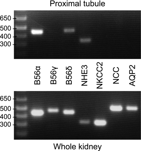
Nephrogenic dopamine is a potent natriuretic paracrine/autocrine hormone that is central for mammalian sodium homeostasis. In the renal proximal tubule, dopamine induces natriuresis partly via inhibition of the sodium/proton exchanger NHE3. The signal transduction pathways and mechanisms by which dopamine inhibits NHE3 are complex and incompletely understood. This manuscript describes the role of the serine/threonine protein phosphatase 2A (PP2A) in the regulation of NHE3 by dopamine. The PP2A regulatory subunit B56δ (coded by the Ppp2r5d gene) directly associates with more than one region of the carboxy-terminal hydrophilic putative cytoplasmic domain of NHE3 (NHE3-cyto), as demonstrated by yeast-two-hybrid, coimmunoprecipitation, blot overlay, and in vitro pull-down assays. Phosphorylated NHE3-cyto is a substrate for purified PP2A in an in vitro dephosphorylation reaction. In cultured renal cells, inhibition of PP2A by either okadaic acid or by overexpression of the simian virus 40 (SV40) small T antigen blocks the ability of dopamine to inhibit NHE3 activity and to reduce surface NHE3 protein. Dopamine-induced NHE3 redistribution is also blocked by okadaic acid ex vivo in rat kidney cortical slices. These studies demonstrate that PP2A is an integral and critical participant in the signal transduction pathway between dopamine receptor activation and NHE3 inhibition.


Figures

Fig. 1.
Fig. 1.
Interaction between Na+/H+ exchanger NHE3 fragments and protein phosphatase 2A regulatory subunit PP2A-B56δ in yeast. Different truncations of the NHE3 C-terminal cytoplasmic tail (NHE3-cyto) were used as baits in a 2-hybrid assay against full-length PP2A-B56δ prey. The NHE3-cyto truncations are represented as horizontal bars labeled with the corresponding amino acids of opossum NHE3. Interaction is expressed semiquantitatively as β-galactosidase activity.
Fig. 2.
Fig. 2.
PP2A-B56δ is expressed in the proximal tubule. RNA was prepared from microdissected rat tubule segments and from whole kidney, reverse-transcribed, and PCR-amplified with primer sets specific for the indicated transcripts. PP2A-B56δ and PP2A-B56α, but not PP2A-B56γ, are expressed in the proximal tubule. NKCC2, Na+-K+-2Cl cotransporter; NCC, Na+-Cl cotransporter; AQP2, aquaporin-2.
Fig. 3.
Fig. 3.
Interaction between NHE3 and PP2A-B56δ in renal cells. A: confluent quiescent opossum kidney Opossum kidney proximal tubule-like (OKP) cells were lysed, and immunoprecipitations (IP) were performed with anti-NHE3 antibody, anti-PP2A-B56δ, or preimmune control antiserum (IgG), followed by SDS-PAGE and immunoblotting (IB) with anti-NHE3 antibody. Native NHE3 and PP2A-B56δ coimmunoprecipitate. B: OKP cells were cotransfected with hemagglutinin (HA)-tagged NHE3 and with Flag-tagged PP2A-B56 subunits (α, γ, or δ), and the anti-HA immune complexes were tested for the presence of the Flag tag. Transfected NHE3 coimmunoprecipitates with transfected PP2A-B56δ and PP2A-B56γ, but not with PP2A-B56α. C: OKP cells were cotransfected with Flag-tagged PP2A-B56δ and myc-tagged NHE3, either full length (FL) or different C-terminus truncations. Full-length NHE3 and the C-terminus truncations Δ775, Δ640, Δ552, but not the truncation Δ462 coimmunoprecipitate with PP2A-B56δ.
Fig. 4.
Fig. 4.
Interaction between NHE3 and PP2A-B56δ in vitro. A: purified 6His-tagged NHE3-cyto and 6His-tagged PP2A-B56δ (baculovirus/Sf9 insect cell expressed) were mixed in vitro, and immunoprecipitations (IP) were performed with either anti-PP2A-B56δ or anti-NHE3. Immunoblotting (IB) was performed with the antibodies indicated. B: PP2A-B56δ purified from Sf9 insect cells was resolved by SDS-PAGE and transferred to nitrocellulose membranes. An overlay was performed with individual fragments of HA-tagged NHE3-cyto, followed by immunoblotting with anti-HA.
Fig. 5.
Fig. 5.
NHE3 is a substrate for PP2A phosphatase activity in vitro. NHE3-cyto was hyperphosphorylated in vitro with the catalytic subunit of PKA and [32P]ATP and subjected to dephosphorylation with purified PP2A for the indicated times. Reactions were terminated by the addition of 2× SDS-sample buffer, resolved by SDS-PAGE, and visualized by phosphorimaging. NHE3-cyto antigen was visualized by immunoblotting.
Fig. 6.
Fig. 6.
PP2A mediates the dopamine-induced inhibition of NHE3 activity in cell culture. Effect of dopamine (10−5 M; 30 min) on NHE3 activity in the presence of the PP2A inhibitors okadaic acid (10−8 M; A) or small T antigen (transient transfection 4 days prior to experiment; B) is shown. Results are presented as means and SD from 4 (A) and 3 (B) experiments, with each condition in each experiment examined in triplicate. *P < 0.01 by ANOVA.
Fig. 7.
Fig. 7.
PP2A mediates the dopamine-induced internalization of NHE3 in cell culture. Effect of dopamine (10−5 M; 40 min) and okadaic acid (10−8 M) on cell surface NHE3 obtained by biotin labeling and streptavidin-agarose affinity precipitation (A) and total cell NHE3 (B) is shown. Results are presented as means and SD from 4 experiments. *P < 0.01 by ANOVA. C: representative immunoblots. D: effect of dopamine (10−5 M; 55 min) and okadaic acid (10−8 M) on native NHE3 in OKP cells by confocal fluorescence microscopy. The figure shows 2 different perspectives (xy and xz planes) for each condition. The apical membrane of the cells is toward the lower part of the xz cross sections (arrows).
Fig. 8.
Fig. 8.
PP2A is important for dopamine-induced NHE3 redistribution in the kidney. Rat cortical kidney slices were incubated for 40 min with vehicle (A), dopamine (10−5 M; B), okadaic acid (10−8 M; C), or dopamine (10−5 M) and okadaic acid (10−8 M; D), stained for NHE3 (red) and actin (green) and visualized by confocal fluorescence microscopy. In the red channel, note the distribution of NHE3 at the tubular lumen in A, C, and D, but not in B. Preserved colocalization with actin (brush border marker) in B may indicate redistribution of NHE3 from the tip to the base of the microvili.

Similar articles

Cited by

References

    1. Albrecht FE, Drago J, Felder RA, Printz MP, Eisner GM, Robillard JE, Sibley DR, Westphal HJ, Jose PA. Role of the D1A dopamine receptor in the pathogenesis of genetic hypertension. J Clin Invest 97: 2283–2288, 1996 - PMC - PubMed
    1. Altschul SF, Gish W, Miller W, Myers EW, Lipman DJ. Basic local alignment search tool. J Mol Biol 215: 403–410, 1990 - PubMed
    1. Amemiya M, Loffing J, Lotscher M, Kaissling B, Alpern RJ, Moe OW. Expression of NHE-3 in the apical membrane of rat renal proximal tubule and thick ascending limb. Kidney Int 48: 1206–1215, 1995 - PubMed
    1. Aperia A, Bertorello A, Seri I. Dopamine causes inhibition of Na+-K+-ATPase activity in rat proximal convoluted tubule segments. Am J Physiol Renal Fluid Electrolyte Physiol 252: F39–F45, 1987 - PubMed
    1. Aperia AC. Intrarenal dopamine: a key signal in the interactive regulation of sodium metabolism. Annu Rev Physiol 62: 621–647, 2000 - PubMed

Publication types

MeSH terms

LinkOut - more resources