. 2010 May;84(9):4673-81.
doi: 10.1128/JVI.02306-09. Epub 2010 Feb 24.

The lack of an inherent membrane targeting signal is responsible for the failure of the matrix (M1) protein of influenza A virus to bud into virus-like particles

Affiliations

The lack of an inherent membrane targeting signal is responsible for the failure of the matrix (M1) protein of influenza A virus to bud into virus-like particles

Dan Wang et al. J Virol. 2010 May.

Abstract

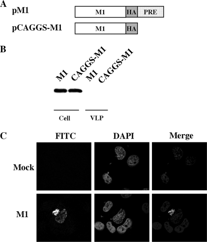
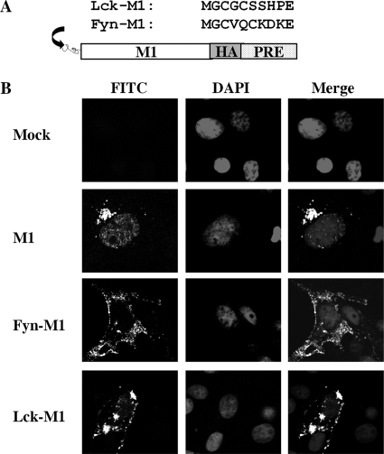
The matrix protein (M1) of influenza A virus is generally viewed as a key orchestrator in the release of influenza virions from the plasma membrane during infection. In contrast to this model, recent studies have indicated that influenza virus requires expression of the envelope proteins for budding of intracellular M1 into virus particles. Here we explored the mechanisms that control M1 budding. Similarly to previous studies, we found that M1 by itself fails to form virus-like-particles (VLPs). We further demonstrated that M1, in the absence of other viral proteins, was preferentially targeted to the nucleus/perinuclear region rather than to the plasma membrane, where influenza virions bud. Remarkably, we showed that a 10-residue membrane targeting peptide from either the Fyn or Lck oncoprotein appended to M1 at the N terminus redirected M1 to the plasma membrane and allowed M1 particle budding without additional viral envelope proteins. To further identify a functional link between plasma membrane targeting and VLP formation, we took advantage of the fact that M1 can interact with M2, unless the cytoplasmic tail is absent. Notably, native M2 but not mutant M2 effectively targeted M1 to the plasma membrane and produced extracellular M1 VLPs. Our results suggest that influenza virus M1 may not possess an inherent membrane targeting signal. Thus, the lack of efficient plasma membrane targeting is responsible for the failure of M1 in budding. This study highlights the fact that interactions of M1 with viral envelope proteins are essential to direct M1 to the plasma membrane for influenza virus particle release.


Figures

FIG. 1.
FIG. 1.
M1 alone is not sufficient for extracellular VLP formation. (A) Schematic diagram of M1 expression constructs. An HA epitope tag (YPYDVPDYA) was appended in frame to the C terminus of the M1 protein in pPRE and pCAGGS vectors, producing pM1 and pCAGGS-M1, respectively. (B) VLP production by COS-1 cells transfected with pM1 and pCAGGS-M1 plasmids. VLPs and cell lysates were analyzed by Western blotting with an anti-HA monoclonal antibody. (C) Representative M1 subcellular localization images in transfected COS-1 cells expressing M1 protein. At 48 h posttransfection, COS-1 cells transfected with either pM1 or empty vector (mock) plasmids were fixed, permeabilized, and stained with goat anti-M1 IgG-FITC. Nuclei were stained with DAPI. M1 localization was then examined using confocal microscopy at an ×60 magnification.
FIG. 2.
FIG. 2.
Comparative studies of M1 localization patterns between transient transfection and virus infection in MDCK cells. (A) Schematic representation of M1 BiFC constructs (M1-VN and M1-VC). Venus fragments VN (N-terminal 173 residues) and VC (C-terminal 83 residues) were fused in frame to the C terminus of M1, which resulted in pM1-VN and pM1-VC, respectively. “L” indicates a linker sequence (YPYDVPDYAASDIAAA) inserted between VN or VC fragments and M1. A portion of the linker is an HA tag sequence. (B) M1 subcellular localization images in MDCK cells expressing BiFC M1-VN and M1-VC fusion proteins. At 48 h posttransfection, MDCK cells transfected with BiFC M1-VN/M1-VC plasmids or M1-VC plasmids (negative control) were fixed, stained with DAPI, and imaged with a confocal microscope at an ×60 magnification. Two representative BiFC images of M1 localization are shown (M1-M1a and M1-M1b). (C) M1 subcellular localization images of virus-infected MDCK cells. At 48 h postinfection, MDCK cells infected with influenza H1N1 A/WSN/33 virus were fixed, permeabilized, and stained with goat anti-M1 IgG-FITC. Sytox orange was used for staining nuclei of infected cells. M1 localization was examined with a confocal microscopy at an ×100 magnification.
FIG. 3.
FIG. 3.
Membrane targeting domains redirect M1 to the plasma membrane. (A) Schematic representation of two membrane targeting domains derived from Fyn and Lck oncoproteins that were appended to the M1 N terminus. (B) Representative M1 subcellular localization images in COS-1 cells expressing M1 or Fyn-M1 or Lck-M1. At 48 h posttransfection, COS-1 cells individually transfected with pM1, pFyn-M1, pLck-M1, and empty vector (mock) were fixed, permeabilized, and stained with goat anti-M1 IgG-FITC. Nuclei of COS-1 cells were stained with DAPI. M1 localization among these cells was then examined by confocal microscopy at an ×60 magnification.
FIG. 4.
FIG. 4.
Production of extracellular M1 particles by membrane-targeted M1 proteins. (A) VLP production by COS-1 cells transfected with pM1 or pFyn-M1 or pLck-M1 plasmid. M1 VLPs and cell lysates were analyzed in a Western blot assay with anti-M1 monoclonal antibody. (B) VLP production by COS-1 cells transfected with Fyn-M1 or FynSS-M1 mutant defective in protein palmitoylation. VLPs and cell lysates were analyzed by Western blotting with anti-M1 monoclonal antibody. (C) VLP production by COS-1 cells transfected with Fyn-M1 in the absence or presence of protein palmitoylation inhibitors, 2-bromopalmitate (2BP; Sigma-Aldrich) and cerulenin (Sigma-Aldrich). VLPs and cell lysates were analyzed by Western blotting with anti-HA monoclonal antibody (Sigma-Aldrich). Cell lysates derived from the 2BP treatment and the DMSO control were also analyzed for the expression of endogenous β-actin proteins with antiactin monoclonal antibody (Abcam). (D) Trypsin digestion of VLPs. Purified VLPs derived from COS-1 cells transfected with Fyn-M1 were treated with TPCK-trypsin (Sigma-Aldrich) in the presence or absence of 1% Triton X-100 for 30 min at 37°C. Fyn-M1 VLPs were also treated with 1% Triton X-100 without TPCK-trypsin under the same experimental conditions. M1 protein was then visualized by Western blotting with anti-HA monoclonal antibody. (E) Representative M1 subcellular localization images in COS-1 cells expressing Fyn-M1 with or without 2BP treatment or Fyn-M1SS. At 48 h posttransfection, COS-1 cells individually transfected with pFyn-M1 (in the presence or absence of 2BP) and pFyn-M1SS were fixed, permeabilized, and stained with goat anti-M1 IgG-FITC. Nuclei of COS-1 cells were stained with DAPI. M1 localization among these cells was then examined by confocal microscopy at an ×60 magnification. (F) Representative GFP subcellular localization images in COS-1 cells expressing Fyn-GFP and Lck-GFP and VLP production by COS-1 cells transfected with pFyn-GFP or pLck-GFP plasmid. GFP VLPs and cell lysates were analyzed in a Western blot assay with anti-GFP polyclonal antibody (Abcam).
FIG. 5.
FIG. 5.
Coexpression of M2 shifts M1 to the plasma membrane and allows M1 particle budding. (A) A schematic diagram of the expression constructs of M2 (pM2) and M2 mutant (pM2ΔCT) lacking the entire CT domain as well as our BiFC M2-VC fusion construct. The experimental approach for the generation of these constructs has been detailed in Materials and Methods. (B) VLP production by COS-1 cells transfected with pM1 plasmid, either alone or in combination with pM2 or pM2ΔCT. At 72 h following transfection, supernatant medium and cellular lysates from each culture were individually collected and VLPs were prepared by ultracentrifugation. Equal amounts of VLP and lysate samples were run on different gels with the same concentration (12.5% SDS-PAGE) under similar experimental conditions, followed by transferring of proteins to different nitrocellulose membranes. Membranes were then probed separately with M1- or M2-specific antibody and exposed to different films for the visualization of protein expression. Note that Ml and M2/M2CTΔ protein bands were placed together arbitrarily according to their molecular weights by comparison with protein markers. Similar results were also obtained in experiments involving the separation of equal amounts of VLPs and cellular lysates on the same gel/membrane followed by cutting of the membrane into two different pieces corresponding to the migration distances of Ml and M2/M2CTΔ proteins according to the position of molecular weight markers. The separated membrane pieces were then individually analyzed in Western blotting via protein-specific antibodies (Ml and M2), respectively. (C) Subcellular localization patterns of M1 protein in transfected COS-1 cells expressing M1, M1 and M2, and M1 and M2ΔCT and subcellular localization patterns of M1-M2 complex in transfected COS-1 cells expressing M1-VN and M1-VC. At 48 h posttransfection, COS-1 cells were fixed, permeabilized, and stained with goat anti-M1 IgG-FITC. For the BiFC experiment, staining with anti-M1 IgG-FITC was not needed. Nuclei of COS-1 cells were stained with DAPI. Two representative images of M1 localization, M1+M2a and M1+M2b, are shown from cells coexpressing M1 and M2 proteins.

Similar articles

Cited by

References

    1. Ali, A., R. T. Avalos, E. Ponimaskin, and D. P. Nayak. 2000. Influenza virus assembly: effect of influenza virus glycoproteins on the membrane association of M1 protein. J. Virol. 74:8709-8719. - PMC - PubMed
    1. Bourmakina, S. V., and A. Garcia-Sastre. 2003. Reverse genetics studies on the filamentous morphology of influenza A virus. J. Gen. Virol. 84:517-527. - PubMed
    1. Burleigh, L. M., L. J. Calder, J. J. Skehel, and D. A. Steinhauer. 2005. Influenza A viruses with mutations in the M1 helix six domain display a wide variety of morphological phenotypes. J. Virol. 79:1262-1270. - PMC - PubMed
    1. Chazal, N., and D. Gerlier. 2003. Virus entry, assembly, budding, and membrane rafts. Microbiol. Mol. Biol. Rev. 67:226-237. - PMC - PubMed
    1. Chen, B. J., G. P. Leser, D. Jackson, and R. A. Lamb. 2008. The influenza virus M2 protein cytoplasmic tail interacts with the M1 protein and influences virus assembly at the site of virus budding. J. Virol. 82:10059-10070. - PMC - PubMed

Publication types

LinkOut - more resources