. 2010 May;84(9):4630-45.
doi: 10.1128/JVI.02431-09. Epub 2010 Feb 24.

Downregulation of Cdc2/CDK1 kinase activity induces the synthesis of noninfectious human papillomavirus type 31b virions in organotypic tissues exposed to benzo[a]pyrene

Affiliations

Downregulation of Cdc2/CDK1 kinase activity induces the synthesis of noninfectious human papillomavirus type 31b virions in organotypic tissues exposed to benzo[a]pyrene

Samina Alam et al. J Virol. 2010 May.

Abstract

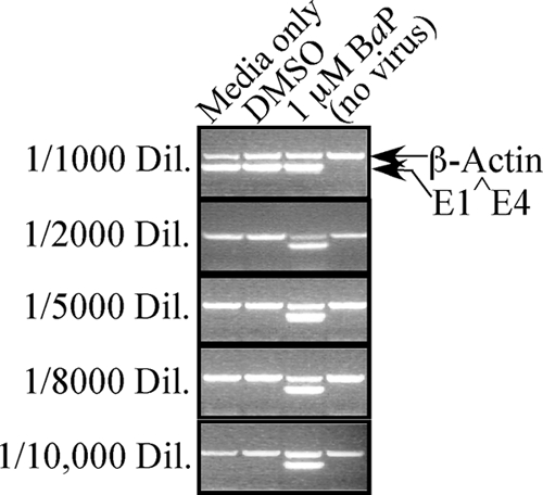
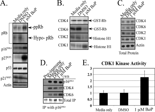
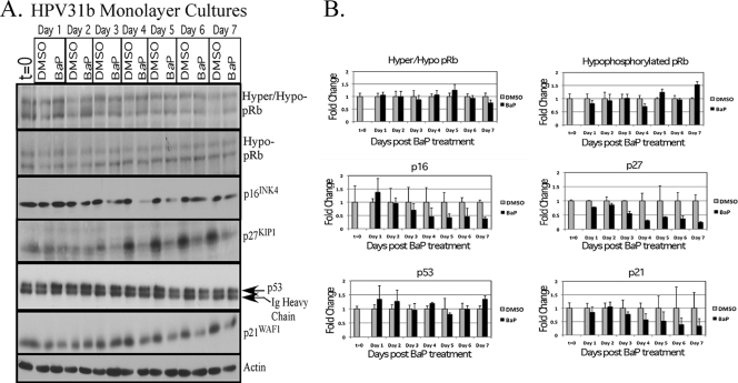
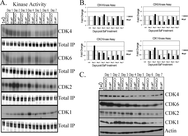
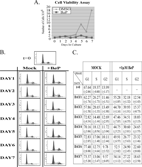
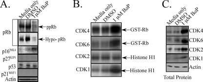
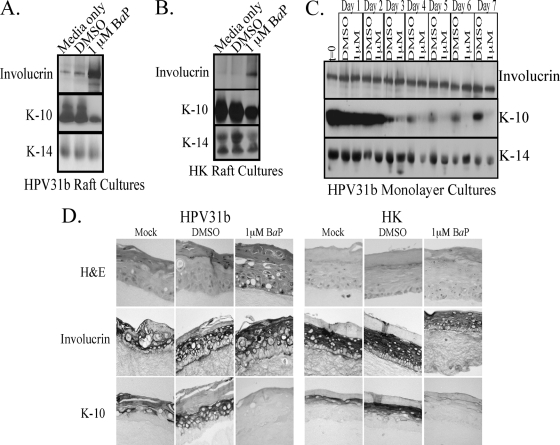
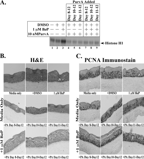
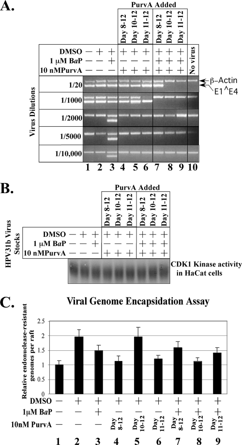
Epidemiological studies suggest that human papillomavirus (HPV)-infected women who smoke face an increased risk for developing cervical cancer. We have previously reported that exposure of HPV-positive organotypic cultures to benzo[a]pyrene (BaP), a major carcinogen in cigarette smoke, resulted in enhanced viral titers. Since BaP is known to deregulate multiple pathways of cellular proliferation, enhanced virion synthesis could result from carcinogen/host cell interaction. Here, we report that BaP-mediated upregulation of virus synthesis is correlated to an altered balance between cell cycle-specific cyclin-dependent kinase (CDK) activity profile compared with controls. Specifically, BaP treatment increased accumulation of hyperphosphorylated retinoblastoma protein (pRb) which coincided with increased cdc2/CDK1 kinase activity, but which further conflicted with the simultaneous upregulation of CDK inhibitors p16(INK4) and p27(KIP1), which normally mediate pRb hypophosphorylation. In contrast, p21(WAF1) and p53 levels remained unchanged. Under these conditions, CDK6 and CDK2 kinase activities were decreased, whereas CDK4 kinase activity remained unchanged. The addition of purvalanol A, a specific inhibitor of CDK1 kinase, to BaP-treated cultures, resulted in the production of noninfectious HPV type 31b (HPV31b) particles. In contrast, infectivity of control virus was unaffected by purvalanol A treatment. BaP targeting of CDK1 occurred independently of HPV status, since BaP treatment also increased CDK1 activity in tissues derived from primary keratinocytes. Our data indicate that HPV31b virions synthesized in the presence of BaP were dependent on BaP-mediated alteration in CDK1 kinase activity for maintaining their infectivity.


Figures

FIG. 1.
FIG. 1.
Infectious titers of HPV31b control raft tissues and tissues treated with 1 μM BaP for 12 days. Shown is a 2% agarose gel of nested reverse transcription-PCR (RT-PCR)-amplified HPV31 E1^E4 spliced transcript and β-actin in reactions with increasing dilutions (Dil.) of the virus stocks shown on the left. Infectivity assays were performed using HaCaT cells as previously described (2). The no-virus control consisted of HaCaT cells incubated with medium only.
FIG. 2.
FIG. 2.
Expression of cell cycle tumor suppressors and an altered profile of CDK kinase activities in differentiating 12-day CIN-612 9E raft cultures correlates with increased HPV31b viral titers. (A) Western blot analysis to detect pRb, p16INK4, p27KIP1, and p21WAF1 from whole-cell extracts prepared from 12-day BaP-treated and control raft cultures. The pRb protein was detected using two different antibodies: rabbit polyclonal antibody (Santa Cruz) detects both the hyperphosphorylated form (ppRb) and hypophosphorylated form (Hypo-pRb) of this protein, whereas the mouse monoclonal antibody (Becton Dickinson) detects exclusively the hypophosphorylated (Hypo-pRb) form. The p16INK4, p27KIP1, and p21WAF1 tumor suppressors were detected using antibodies as described in Materials and Methods. The p53 protein was immunoprecipitated from raft tissues and detected using Western blot analysis as described in Materials and Methods and reference . (B) Kinase assays were performed to detect CDK4 and CDK6 activities using GST-Rb as the substrate and CDK1- and CDK2-associated kinase activities using histone H1 as the substrate. HPV31b raft cell extracts were prepared from BaP-treated and control tissues followed by determination of kinase activities as indicated. (C) Western blot analysis was performed to detect the total levels of CDK4, CDK6, CDK2, and CDK1 protein in 1 μM BaP-treated and control HPV31b raft tissues. (D) Immunoprecipitation of p16INK4 and detection of coprecipitating levels of bound CDK4 and CDK6. Control HPV31b raft tissues and tissues treated with 1 μM BaP were used to prepare protein extracts, followed by immunoprecipitation (IP) of the p16INK4 protein. For determining quantitative binding of CDK4 and CDK6 to p16INK4 protein, the levels of immunoprecipitated p16INK4 were normalized using densitometric analysis. On the basis of the normalized levels, Western blotting (WB) was performed using antibody to p16INK4. The p16INK4 immunoprecipitates were further used in Western blots to detect coprecipitated CDK4 and CDK6 using the antibodies described above. Results shown are representative of three individual experiments. (E) Comparative densitometric analysis of the bands depicting CDK1-associated kinase activity in 1 μM BaP-treated and control raft tissues. The medium-only control was set at 1. The y axis shows CDK1 kinase activity (fold change). The values are the averages plus standard deviations of the means (error bars) for three independent experiments.
FIG. 3.
FIG. 3.
Expression of cell cycle tumor suppressors in nondifferentiating monolayer cultures. (A) Western blot analysis to detect pRb, p16INK4, p27KIP1, and p21WAF1 from whole-cell extracts prepared from 1 μM BaP-treated and control DMSO-treated monolayer cultures over a 7-day period. The cells were synchronized by trypsinization of 80% confluent monolayer cultures and plating at a density of 1 × 10⁶ cells in E medium. The cells were incubated for 10 to 12 h at which point two-thirds of the cells are maximally synchronized in the G1 phase. This time point is designated time zero (t = 0). The 1 μM BaP and DMSO treatments were added at this point. Cells were trypsinized and pelleted over a period of 7 days. Cells were passaged at a ratio of 1:2 on day 2 and day 5. The pRb protein was detected using two different antibodies: rabbit polyclonal antibody (Santa Cruz) detects both the hyperphosphorylated (ppRb) and hypophosphorylated (Hypo-pRb) forms of this protein, whereas the mouse monoclonal antibody (Becton Dickinson) detects exclusively the hypophosphorylated form (Hypo-pRb). The p16INK4, p27KIP1, and p21WAF1 tumor suppressors were detected using antibodies as described in Materials and Methods. The p53 protein was immunoprecipitated from monolayer cultures and detected using Western blot analysis as described in Materials and Methods and reference . (B) Comparative densitometric analysis of the protein bands in Western blots depicting expression of pRb, p16INK4, p27KIP1, and p21WAF1 proteins in 1 μM BaP-treated and control HPV31b monolayer cultures. The medium-only control was set at 1. The values are the averages plus standard deviations of the means (error bars) for three independent experiments.
FIG. 4.
FIG. 4.
Determination of CDK-associated kinase activities in BaP-treated and control HPV31b monolayer cultures. The cells were synchronized by trypsinization of 80% confluent monolayer cultures and plating at a density of 1 × 10⁶ cells in E medium. The cells were further incubated for 10 to 12 h at which point two-thirds of the cells are maximally synchronized in the G1 phase. This time point is designated time zero (t = 0). The 1 μM BaP and DMSO treatments were added at this point. The cells were treated with 1 μM BaP or treated with DMSO as a control over a 7-day period. The cells were passaged 1:2 on day 2 and day 5. (A) Kinase assays were performed to detect CDK4, CDK6-associated kinase activities using GST-Rb as the substrate, and CDK1- and CDK2-associated kinase activities using histone H1 as the substrate. IP, immunoprecipitation. (B) Comparative densitometric analysis of the bands in kinase assay blots depicting kinase activity of CDK4, CDK6, CDK2, and CDK1 in 1 μM BaP-treated and control HPV31b monolayer cultures. The medium-only control was set at 1. The results from one experiment are shown but are representative results. (C) Western blot analysis was performed to detect total protein levels of CDK4, CDK6, CDK2, and CDK1 in 1 μM BaP-treated and control HPV31b monolayer cultures as described in Materials and Methods.
FIG. 5.
FIG. 5.
Cell cycle progression in 1 μM BaP-treated HPV31b monolayer cultures. Cells were synchronized by trypsinization of 80% confluent monolayer cultures and plating at a density of 1 × 10⁶ cells in E medium. The cells were incubated for 10 to 12 h at which point two-thirds of the cells are maximally synchronized in the G1 phase. This time point is designated time zero (t = 0). The 1 μM BaP and DMSO treatments were added at this point, and cells were treated over a 7-day period. Cells were passed 1:2 on day 2 and day 5. (A) Cell viability assay. Trypan blue exclusion was used to determine the growth inhibitory effect of 1 μM BaP on HPV31 monolayer cultures. Results shown are representative of three individual experiments. (B) Fluorescence-activated cell sorting (FACS) analysis. FACS analysis was performed to determine the percentage of cells in G1, S, and G2/M phases of the cell cycle in 1 μM BaP-treated and control HPV31b monolayer cultures. CIN-612 9E cells were synchronized and treated with BaP or mock treated with DMSO. On each day, cells were harvested by trypsinization, washed with PBS, fixed in 70% ethanol, and stored at −20°C, followed by resuspending the cells in PBS containing 0.1% Triton X-100, 200 μg/ml DNase-free RNase A, and 100 μg/ml of propidium iodide for 30 min at 37°C. Flow cytometric analysis of 106 cells was carried out in a fluorescence-activated cell sorter, and the percentages of cells in the G1, S, and G2/M phases of the cell cycle were determined using the Cell Quest program of Becton Dickinson. Data were analyzed with the Mod Fit LT program. (C) FACS analyses were repeated three times. Results shown represent averages determined from three individual experiments (−, no data), with standard deviations presented in parentheses.
FIG. 6.
FIG. 6.
Expression of cell cycle tumor suppressors and an altered profile of CDK kinase activities in differentiating 12-day HK raft cultures. (A) Western blot analysis to detect pRb, p16INK4, p27KIP1, and p21WAF1 from whole-cell extracts prepared from 1 μM BaP-treated and control raft cultures. The pRb protein was detected using two different antibodies: rabbit polyclonal antibody (Santa Cruz) detects both the hyperphosphorylated (ppRb) and hypophosphorylated (Hypo-pRb) forms of this protein, whereas the mouse monoclonal antibody (Becton Dickinson) detects exclusively the hypophosphorylated form. The p16INK4, p27KIP1, and p21WAF1 tumor suppressors were detected using antibodies as described in Materials and Methods. The p53 protein was immunoprecipitated from raft tissues and detected using Western blot analysis as described in Materials and Methods and reference . (B) Kinase assays were performed to detect CDK4 and CDK6 activities using GST-Rb as the substrate and CDK1- and CDK2-associated kinase activities using histone H1 as the substrate. HK raft cell extracts were prepared from BaP-treated and control tissues followed by determination of kinase activities as indicated. (C) Western blot analysis was performed to detect total levels of CDK4, CDK6, CDK2, and CDK1 protein in 1 μM BaP-treated and control HK raft tissues. Results shown are representative of three individual experiments.
FIG. 7.
FIG. 7.
Expression of keratinocyte differentiation markers. (A and B) Western blot analysis to determine expression of differentiation markers involucrin, K-10, and K-14 from whole-cell extracts prepared from 12-day BaP-treated and control tissues from HPV31b raft cultures (A) and HK raft cultures (B). A total of 5 μg of total protein was used for detecting each protein using Western blot analysis as described in Materials and Methods. (C) Western blot analysis to detect involucrin, K-10, and K-14 from whole-cell extracts prepared from BaP-treated and control HPV31b monolayer cultures. Cells were synchronized by trypsinization of 80% confluent monolayer cultures and plating at a density of 1 × 10⁶ cells in E medium. The cells were incubated for 10 to 12 h at which point two-thirds of the cells are maximally synchronized in the G1 phase. This time point is designated time zero (t = 0). The 1 μM BaP and DMSO treatments were added at this point, and cells were treated over a 7 day period. Cells were passed 1:2 on day 2 and day 5. (D) Immunohistochemical analysis of control and BaP-treated HPV31b (left) and HK (right) raft tissues. (Top row) Hematoxylin and eosin (H&E) staining of raft culture tissue sections treated with control and BaP. (Middle row) Immunohistochemical staining with involucrin antibody. (Bottom row) Immunohistochemical staining with K-10 antibody.
FIG. 8.
FIG. 8.
Effects of purvalanol A on HPV31b infectivity and morphogenesis. (A) 10 nM purvalanol A treatment inhibits CDK1 kinase activity. HPV31b raft tissue extracts were prepared from control and treated tissues. The treated tissues were treated with 1 μM BaP alone for 12 days, treated with 10 nM purvalanol A (PurvA) alone, or cotreated with 1 μM BaP and 10 nM purvalanol A. Kinase activity was determined using histone H1 as the substrate. (B) Purvalanol A treatment does not interfere with tissue differentiation. HPV31b raft tissues treated with 1 μM BaP alone, treated with 10 nM purvalanol A alone (+PA), and cotreated with 1 μM BaP and 10 nM purvalanol A. Treated and control tissues were embedded in paraffin, and thin sections were prepared for H&E staining. (C) Purvalanol A treatment is not toxic to cell growth and nuclear functions. HPV31b raft tissues treated with 1 μM BaP alone or with 10 nM purvalanol A alone or cotreated with 1 μM BaP and 10 nM purvalanol A. Treated and control tissues were embedded in paraffin, and thin sections were stained with an rabbit polyclonal antibody against PCNA. Results shown are representative of three individual experiments.
FIG. 9.
FIG. 9.
Effects of purvalanol A treatment on viral titers. (A) 12-day HPV31b raft tissues treated with 1 μM BaP alone (lane 3) or with 10 nM purvalanol A (PurvA) alone (lanes 4 to 6) or cotreated with 1 μM BaP and 10 nM purvalanol A (lanes 7 to 9) and control tissues (lanes 1 and 2) were used for preparing crude viral stocks followed by infection of HaCaT cells as we have described previously (2). Shown is a 2% agarose gel of nested RT-PCR-amplified HPV31 E1^E4 spliced transcript and β-actin in reaction mixtures with increasing dilutions of the virus stocks shown on the left. (B) Lack of HPV31b infectivity of virions cotreated with BaP and purvalanol A is not a side effect of residual purvalanol A activity. HPV31b virus stocks tested in Fig. 9A were used to infect HaCaT cells followed by 48 h of incubation. Cells were harvested, and then CDK1 kinase activity was detected using histone H1 as the substrate. (C) Lack of HPV31b infectivity of virus cotreated with BaP and purvalanol A was not due to disassembly or lack of assembly of viral particles. HPV31b raft tissues treated with 1 μM BaP alone (bar 3) or 10 nM purvalanol A alone (bars 4 to 6) or cotreated with 1 μM BaP and 10 nM purvalanol A (bars 7 to 9) and control tissues (bars 1 and 2) were used for preparing crude viral stocks which were subsequently treated with benzonase to remove endonuclease-susceptible viral genomes. A qPCR-based assay was used to analyze relative numbers of endonuclease-resistant HPV31b genomes as described in Materials and Methods. Results shown are representative of three individual experiments.

Similar articles

Cited by

References

    1. Abelev, G. I. 2000. Differentiation mechanisms and malignancy. Biochemistry (Mosc.) 65:107-116. - PubMed
    1. Alam, S., M. J. Conway, H. S. Chen, and C. Meyers. 2008. The cigarette smoke carcinogen benzo[a]pyrene enhances human papillomavirus synthesis. J. Virol. 82:1053-1058. - PMC - PubMed
    1. Alam, S., E. Sen, H. Brashear, and C. Meyers. 2006. Adeno-associated virus type 2 increases proteosome-dependent degradation of p21WAF1 in a human papillomavirus type 31b-positive cervical carcinoma line. J. Virol. 80:4927-4939. - PMC - PubMed
    1. Andrysik, Z., J. Vondracek, M. Machala, P. Krcmar, L. Svihalkova-Sindlerova, A. Kranz, C. Weiss, D. Faust, A. Kozubik, and C. Dietrich. 2007. The aryl hydrocarbon receptor-dependent deregulation of cell cycle control induced by polycyclic aromatic hydrocarbons in rat liver epithelial cells. Mutat. Res. 615:87-97. - PubMed
    1. Asselineau, D., and M. Prunieras. 1984. Reconstruction of ‘simplified’ skin: control of fabrication. Br. J. Dermatol. 111(Suppl. 27):219-222. - PubMed

Publication types