. 2010 May;84(9):4289-301.
doi: 10.1128/JVI.02534-09. Epub 2010 Feb 24.

Avian reovirus microNS protein forms homo-oligomeric inclusions in a microtubule-independent fashion, which involves specific regions of its C-terminal domain

Affiliations

Avian reovirus microNS protein forms homo-oligomeric inclusions in a microtubule-independent fashion, which involves specific regions of its C-terminal domain

Alberto Brandariz-Nuñez et al. J Virol. 2010 May.

Abstract

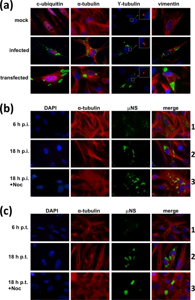
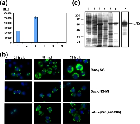
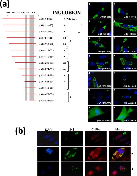
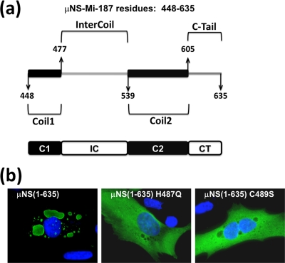
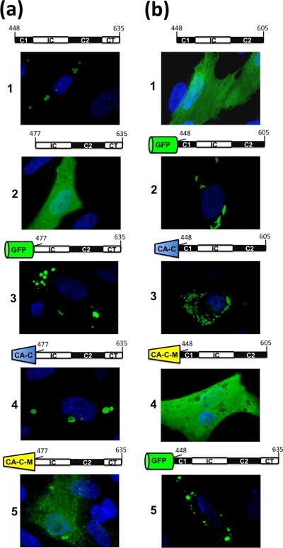
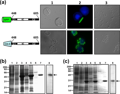
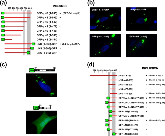
Members of the genus Orthoreovirus replicate in cytoplasmic inclusions termed viral factories. Compelling evidence suggests that the nonstructural protein microNS forms the matrix of the factories and recruits specific viral proteins to these structures. In the first part of this study, we analyzed the properties of avian reovirus factories and microNS-derived inclusions and found that they are nonaggresome cytoplasmic globular structures not associated with the cytoskeleton which do not require an intact microtubule network for formation and maturation. We next investigated the capacity of avian reovirus microNS to form inclusions in transfected and baculovirus-infected cells. Our results showed that microNS is the main component of the inclusions formed by recombinant baculovirus expression. This, and the fact that microNS is able to self-associate inside the cell, suggests that microNS monomers contain all the interacting domains required for inclusion formation. Examination of the inclusion-forming capacities of truncated microNS versions allowed us to identify the region spanning residues 448 to 635 of microNS as the smallest that was inclusion competent, although residues within the region 140 to 380 seem to be involved in inclusion maturation. Finally, we investigated the roles that four different motifs present in microNS(448-635) play in inclusion formation, and the results suggest that the C-terminal tail domain is a key determinant in dictating the initial orientation of monomer-to-monomer contacts to form basal oligomers that control inclusion shape and inclusion-forming efficiency. Our results contribute to an understanding of the generation of structured protein aggregates that escape the cellular mechanisms of protein recycling.


Figures

FIG. 1.
FIG. 1.
Relationship of ARV factories and μNS-derived inclusions with the cytoskeleton and aggresome generation. (a) Semiconfluent CEF monolayers were mock infected (top row), infected with 5 PFU/cell of avian reovirus S1133 (middle row), or transfected with 1 μg of pCINeo-μNS(S1133) per well (bottom row) and then fixed at 18 h p.i. or 18 h p.t. The cells were immunostained with rabbit anti-μNS serum and then with Alexa 488-conjugated goat anti-rabbit IgG (green) and counterstained with mouse monoclonal antibodies to either conjugated ubiquitin, α-tubulin, γ-tubulin, or vimentin, as indicated above each column, followed by Alexa 594-conjugated goat anti-mouse IgG (red). Nuclei were stained with DAPI (blue). The γ-tubulin-stained MTOC is amplified for clarity. The insets are enlargements of the boxed areas. (b) Semiconfluent monolayers of CEFs were infected with 5 PFU/cell of avian reovirus S1133, and the cells were either untreated (6 h p.i. and 18 h p.i.) or incubated with 10 μM nocodazole from 0 to 18 h postinfection (18 h p.i.+Noc). All cells were fixed at the times postinfection indicated on the left and immunostained using rabbit anti-μNS serum and Alexa 488-conjugated goat anti-rabbit IgG (green). Microtubules were visualized by immunostaining them with mouse monoclonal antibodies to α-tubulin, followed by Alexa 594-conjugated goat anti-mouse IgG (red). Nuclei were counterstained with DAPI (blue). (c) Same as panel b, but the CEFs were transfected with μNS-expressing plasmid instead of infected. On the left are indicated the hours posttransfection when the cells were fixed, either untreated (6 h p.t. and 18 h p.t.) or treated with 10 μM nocodazole from 0 to 18 h posttransfection (18 h p.t.+Noc).
FIG. 2.
FIG. 2.
Mammalian two-hybrid analysis, and baculovirus expression of μNS-derived constructs in insect cells. (a) Mammalian Matchmaker two-hybrid analysis of μNS self-interaction. The bars represent the luciferase levels of extracts from Cos-7 cells transfected with the following plasmid combinations: 1, positive control included in the system (see Materials and Methods); 2, empty plasmids expressing activation and DNA-binding domains plus the luciferase reporter plasmid; 3, plasmids expressing μNS fused to the activation and DNA-binding domains plus luciferase reporter plasmid; 4, plasmids expressing μNS fused to the activation domain and empty plasmid expressing the DNA-binding domain plus luciferase reporter plasmid; 5, empty plasmid expressing the activation domain and plasmid expressing μNS fused to the DNA-binding domain plus luciferase reporter plasmid; 6, untransfected cells. RLU are indicated on the left. The error bars indicate standard deviations. (b) Time course analysis showing the subcellular localization of μNS, μNS-Mi, and CA-C-μNS(448-605) proteins in Sf9 cells. The last two proteins are described in Fig. 5 and 7, but they are included here for comparison. Semiconfluent monolayers of Sf9 cells were infected with a recombinant baculovirus expressing μNS (top), μNS-Mi (middle), or CA-C-μNS(448-605) (bottom). The cells were then fixed and immunostained with rabbit antibodies raised against μNS, followed by Alexa 488-conjugated goat anti-rabbit IgG (green), at the infection times indicated above the images. Nuclei were counterstained with DAPI (blue). (c) Expression, purification, and immunoblot analysis of μNS-derived inclusions. Sf9 insect cells infected with a recombinant baculovirus expressing μNS were lysed in hypotonic buffer at 72 h p.i., and the resulting cell extract (lane 3) was fractionated by centrifugation into pellet and supernatant fractions (the supernatant fraction is shown in lane 4). The pellet was then washed twice with hypotonic buffer, resuspended in the same volume of hypotonic buffer, and sonicated. The sonicated extract (lane 5) was centrifuged, and the pelleted inclusions were washed five times with hypotonic buffer (lane 6). Mock-infected and wild-type-baculovirus-infected Sf9 cell extracts, obtained by lysing the cells in hypotonic buffer at 72 h p.i., are shown in lanes 1 and 2, respectively. All samples were resolved by 10% SDS-PAGE, and the protein bands were visualized by Coomassie blue staining. The position of recombinant μNS is indicated on the right and those of the molecular weight markers on the left. The sample in lane 6 (purified μNS inclusions) was subjected to Western blot analysis with anti-μNS antibodies (lane 7).
FIG. 3.
FIG. 3.
Summary of the expression of C-terminal truncations of μNS. Full-length μNS is schematically indicated by a horizontal red bar comprising residues 1 to 635 (the positions are numbered on the top and right). A similar red bar indicates each single truncation, spanning the approximate region corresponding to the construct. The positions of two previously described coiled-coil elements predicted in the sequence of μNS are indicated by two vertical gray bars. The ability of each construct to form intracellular inclusions is indicated as positive (+) or negative (−), and representative immunofluorescence images of transfected CEFs are presented on the right. The immunofluorescence analysis was performed as indicated in Fig. 2b on CEFs fixed after 18 h of transfection with plasmids expressing the indicated C-terminal μNS truncations.
FIG. 4.
FIG. 4.
Summary of the expression of N-terminal deletions of μNS. (a) N-terminal deletions of μNS are indicated as in Fig. 3. The ability of each construct to form intracellular inclusions is indicated as positive (+), negative (−), or aggregated (ag). The immunofluorescence analysis shown on the right was performed as indicated in Fig. 2b on CEFs fixed after 18 h of transfection with plasmids expressing the indicated N-terminal μNS truncations. (b) Ubiquitination analysis of N-terminal deletions. CEFs were fixed after 18 h of transfection with plasmids expressing μNS(140-635) (row 1), μNS(112-635) (row 2), or μNS(381-635) (row 3) as examples of large, aggregated, and small inclusions, respectively. The cells were immunostained with rabbit anti-μNS antibodies (μNS) and mouse anti-conjugated ubiquitin (C-Ubq). The secondary antibodies used were Alexa 488-conjugated goat anti-rabbit IgG (green) and Alexa 594-conjugated goat anti-mouse IgG (red), respectively. In the merged image, colocalization of μNS(112-635) (row 2) and conjugated ubiquitin is indicated by yellow. Nuclei were counterstained with DAPI (blue).
FIG. 5.
FIG. 5.
Domain composition of μNS-Mi and relevance of the Intercoil domain. (a) Schematic drawing of the minimal μNS region found to retain inclusion-forming ability (μNS-Mi). The four constituent domains are indicated by the amino acid residues that mark the interdomain positions: Coil1 or C1 (448 to 477), Intercoil or IC (477 to 539), Coil2 or C2 (539 to 605), and C-Tail or CT (605 to 635). (b) Immunofluorescence analysis of CEFs transfected with plasmids expressing full-length μNS(1-635) and μNS containing the single-amino-acid Intercoil mutations indicated. The Cells were immunostained as in Fig. 2b.
FIG. 6.
FIG. 6.
Immunofluorescence analysis of the intracellular expression of μNS-Mi chimeras. CEFs were transfected with plasmids expressing μNS-Mi (a1) or the different constructs indicated above each panel. The four domains present in μNS-Mi are represented following the scheme shown in Fig. 5. The green barrel represents green fluorescent protein, whereas blue and yellow truncated cones represent CA-C and CA-C-M, respectively. The transfected cells were stained as in Fig. 2b, except those containing GFP that were visualized without antibodies. (a) Substitution of Coil1. (b) Substitution of C-Tail.
FIG. 7.
FIG. 7.
Baculovirus expression and purification of μNS-Mi chimeras. (a) Sf9 cells were infected with baculoviruses expressing the chimeras indicated on the left. The cells were fixed 3 days after infection and then either treated with DAPI to stain the nuclei (top row) or stained with DAPI and anti-μNS antibodies and photographed under a bright-field (column 1) or fluorescence (column 2) microscope. Column 3 shows images of the tubular (top) or globular (bottom) inclusions purified from the cytoplasm of infected Sf9 cells. (b) Expression, purification, and immunoblot analysis of tubular inclusions (GFP-C1-IC-C2). Sf9 cells infected with baculovirus expressing GFP-C1-IC-C2 were lysed in hypotonic buffer at 72 h p.i., and the resulting cell extract (lane 3) was fractionated by centrifugation into pellet and supernatant fractions (the supernatant fraction is shown in lane 4). The pellet was washed twice with hypotonic buffer, resuspended in the same volume of hypotonic buffer, and sonicated. The sonicated extract (lane 5) was centrifuged and fractionated into pellet and supernatant fractions (the supernatant fraction is shown in lane 6). The pellet was then washed and centrifuged five times with hypotonic buffer (lane 7). Mock-infected or wild-type-baculovirus-infected Sf9 cell extracts lysed in hypotonic buffer at 72 h p.i. are shown in lanes 1 and 2, respectively. All samples were resolved by 12.5% SDS-PAGE, and the protein bands were visualized by Coomassie blue staining. The position of recombinant GFP-C1-IC-C2 is indicated by an arrow on the right and those of the molecular weight markers on the left. The sample in lane 7 (purified tubular inclusions) was subjected to Western blot analysis with anti-μNS antibodies (lane 8). (c) Expression, purification, and immunoblot analysis of globular inclusions (CA-C-C1-IC-C2). The expression and purification of globular inclusions was performed as for panel b. The position of recombinant CA-C-C1-IC-C2 is indicated by an arrow on the right and those of the protein markers on the left. The sample in lane 7 (purified globular inclusions) was subjected to Western blot analysis with anti-μNS antibodies (lane 8).
FIG. 8.
FIG. 8.
Summary of the expression of different μNS-derived constructs. (a and d) Schematic representations of the different constructs. The presence of fused GFP or CA-C is indicated with a green barrel. +, inclusion-forming constructs; −, constructs lacking inclusion-forming activity. Constructions already shown in other figures are indicated in parentheses on the right. (b and c) Fluorescence microscope analysis of CEFs transfected with the indicated plasmids. Nuclei were stained blue with DAPI.

Similar articles

Cited by

References

    1. Attoui, H., F. Billoir, P. Biagini, P. de Micco, and X. de Lamballerie. 2000. Complete sequence determination and genetic analysis of Banna virus and Kadipiro virus: proposal for assignment to a new genus (Seadornavirus) within the family Reoviridae. J. Gen. Virol. 81:1507-1515. - PubMed
    1. Bodelon, G., L. Labrada, J. Martinez-Costas, and J. Benavente. 2001. The avian reovirus genome segment S1 is a functionally tricistronic gene that expresses one structural and two nonstructural proteins in infected cells. Virology 290:181-191. - PubMed
    1. Broering, T. J., M. M. Arnold, C. L. Miller, J. A. Hurt, P. L. Joyce, and M. L. Nibert. 2005. Carboxyl-proximal regions of reovirus nonstructural protein muNS necessary and sufficient for forming factory-like inclusions. J. Virol. 79:6194-6206. - PMC - PubMed
    1. Broering, T. J., J. Kim, C. L. Miller, C. D. Piggott, J. B. Dinoso, M. L. Nibert, and J. S. Parker. 2004. Reovirus nonstructural protein muNS recruits viral core surface proteins and entering core particles to factory-like inclusions. J. Virol. 78:1882-1892. - PMC - PubMed
    1. Broering, T. J., J. S. Parker, P. L. Joyce, J. Kim, and M. L. Nibert. 2002. Mammalian reovirus nonstructural protein microNS forms large inclusions and colocalizes with reovirus microtubule-associated protein micro2 in transfected cells. J. Virol. 76:8285-8297. - PMC - PubMed

Publication types

Substances

LinkOut - more resources