. 2010 May;84(9):4832-9.
doi: 10.1128/JVI.02405-09. Epub 2010 Feb 24.

Pathogenic hantaviruses direct the adherence of quiescent platelets to infected endothelial cells

Affiliations

Pathogenic hantaviruses direct the adherence of quiescent platelets to infected endothelial cells

Irina N Gavrilovskaya et al. J Virol. 2010 May.

Abstract

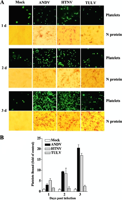
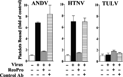
Hantavirus infections are noted for their ability to infect endothelial cells, cause acute thrombocytopenia, and trigger 2 vascular-permeability-based diseases. However, hantavirus infections are not lytic, and the mechanisms by which hantaviruses cause capillary permeability and thrombocytopenia are only partially understood. The role of beta(3) integrins in hemostasis and the inactivation of beta(3) integrin receptors by pathogenic hantaviruses suggest the involvement of hantaviruses in altered platelet and endothelial cell functions that regulate permeability. Here, we determined that pathogenic hantaviruses bind to quiescent platelets via a beta(3) integrin-dependent mechanism. This suggests that platelets may contribute to hantavirus dissemination within infected patients and provides a means by which hantavirus binding to beta(3) integrin receptors prevents platelet activation. The ability of hantaviruses to bind platelets further suggested that cell-associated hantaviruses might recruit platelets to the endothelial cell surface. Our findings indicate that Andes virus (ANDV)- or Hantaan virus (HTNV)-infected endothelial cells specifically direct the adherence of calcein-labeled platelets. In contrast, cells comparably infected with nonpathogenic Tula virus (TULV) failed to recruit platelets to the endothelial cell surface. Platelet adherence was dependent on endothelial cell beta(3) integrins and neutralized by the addition of the anti-beta(3) Fab fragment, c7E3, or specific ANDV- or HTNV-neutralizing antibodies. These findings indicate that pathogenic hantaviruses displayed on the surface of infected endothelial cells bind platelets and that a platelet layer covers the surface of infected endothelial cells. This fundamentally changes the appearance of endothelial cells and has the potential to alter cellular immune responses, platelet activation, and endothelial cell functions that affect vascular permeability. Hantavirus-directed platelet quiescence and recruitment to vast endothelial cell beds further suggests mechanisms by which hantaviruses may cause thrombocytopenia and induce hypoxia. These findings are fundamental to our understanding of pathogenic-hantavirus regulation of endothelial cell responses that contribute to vascular permeability.


Figures

FIG. 1.
FIG. 1.
Pathogenic hantaviruses bind platelets. Platelets were collected in sodium citrate (final concentration, 0.4%) supplemented with 1 μM prostaglandin E1 (PGE1) (Calbiochem) (27), and platelet-rich plasma (PRP) was prepared by centrifugation for 15 min at 1,800 rpm (700 × g) at 25°C. PRP was supplemented with 10 mM EDTA, and platelets were pelleted by centrifugation (15 min at 1,300 × g at 25°C) and washed with HBMT buffer (HEPES-buffered modified Tyrodes buffer; 10 mM HEPES, pH 7.45, 137 mM NaCl, 2.7 mM KCl, 0.4 mM NaH2PO4, 12 mM NaHCO3, 1 mM MgCl2, 0.1% dextrose, 0.2% bovine serum albumin [BSA] with 1 μM PGE1,10 mM EDTA) (27). Platelets were purified over a Sepharose 2B column equilibrated with HBMT at room temperature and quantitated using a hemacytometer (27). Platelets (107 or 108 per ml) were incubated with 104 FFU of HTNV, ANDV, or TULV for 1 h in an incubator at 37°C and 5% CO2. Platelets were pelleted and washed three times with 10 volumes of HBMT, resuspended in 100 μl HBMT, and then adsorbed onto VeroE6 cells in duplicate wells of a 96-well plate (1 h at 37°C). Monolayers were washed with Dulbecco's modified Eagle's medium supplemented with 2% fetal calf serum, and VeroE6 cells were incubated (37°C, 5% CO2) for 24 h prior to methanol fixation (100%, −20°C) (24). The hantavirus nucleocapsid protein present in infected cells was detected by immunoperoxidase staining as previously described, and the number of N protein-containing cells was quantitated (26). Error bars represent the means ± standard deviations (n = 12) of results from four independent experiments.
FIG. 2.
FIG. 2.
Pathogenic-hantavirus binding to platelets is inhibited by β3 integrin antibodies. Platelets (106) were incubated with increasing concentrations of the c7E3(ReoPro) Fab fragment (10) directed at αvβ3 or control antibody (anti-β5 integrin [product no. 1926; Chemicon]) (10 to 100 μg/ml) or mock treated. Subsequently, platelets were incubated with ANDV, HTNV, or TULV (104 FFU) for 2 h at room temperature in HBMT. Following centrifugation and washing 3 times with HBMT to remove unbound virus, platelets were resuspended and adsorbed to VeroE6 cells as described in the legend to Fig. 1. The number of nucleocapsid protein-positive cells is presented as a percentage of the number of mock-treated infected cells (26). Results from two independent experiments are presented.
FIG. 3.
FIG. 3.
Platelets adhere to pathogenic-hantavirus-infected endothelial cells. Human umbilical vein endothelial cells were purchased from Cambrex and seeded onto 96-well plates at a cell density of 2 × 10⁴ cells and grown in endothelial basal medium 2 (Cambrex) containing 10% fetal calf serum, as previously described (25). Confluent endothelial cells were infected in duplicate with HTNV, ANDV, and TULV at an MOI of 0.5 or mock infected. Platelets were purified as described in the legend to Fig. 1 and labeled with calcein AM (5 mM, 15 min, 37°C; Molecular Probes) in HBMT (5, 27). Platelets were pelleted and washed 3 times prior to resuspension in HBMT buffer. At the indicated times postinfection, cells were washed with HBMT (calcium free, with 1 mM MgCl2), incubated with calcein-labeled platelets (107 platelets/well) for 30 min at 37°C, and subsequently washed twice with HBMT buffer. (A) Platelet adherence to endothelial cells was visualized using an Olympus IX51 fluorescence microscope. At least four independent experiments were performed, with similar results. Identically infected endothelial cell monolayers were fixed with methanol, immunoperoxidase stained for the hantavirus N protein, and visualized by light microscopy. (B) The number of platelets bound to endothelial cell monolayers was quantitated using the NIH Image program. Results are presented as fold change relative to level of mock-infected cells. Error bars represent the means ± standard deviations (n = 12) of results from four experiments.
FIG. 4.
FIG. 4.
Neutralizing antibodies to ANDV or HTNV specifically inhibit platelet binding. Endothelial cells were grown in 96-well plates and infected with ANDV, HTNV, or TULV (MOI of 0.5) or mock infected as described in the legend to Fig. 2. At 3 days postinfection, antibodies (Ab) to the ANDV surface glycoproteins (neutralizing titer, 1:640; gift from J. W. Hooper, USAMRIID) (62), neutralizing antibody to HTNV (WHO Reference Center, Seoul, South Korea), or control rabbit serum was added (10 ng/ml) to infected or mock-infected cells for 1 h at 4°C. Subsequently, calcein-labeled platelets (107 platelets/well) were incubated with cells for 30 min at 37°C, and cells were washed three times with HBMT buffer. (A) Platelet adherence to endothelial cells was visualized using an Olympus IX51 fluorescence microscope. (B) Bound platelets were quantified, and results are expressed as fold changes for platelets bound versus the level of the control ± standard deviations for each group. A two-tailed Student t test was used to analyze the statistical differences between the control and treated groups.
FIG. 5.
FIG. 5.
β3 integrin antibodies inhibit platelet adherence to hantavirus-infected endothelial cells. Endothelial cells were infected with ANDV, HTNV, or TULV (MOI of 0.5) or mock infected as described in the legend to Fig. 2. At 3 days postinfection, cells were pretreated with c7E3(ReoPro) antibody (100 ng/ml) (10) or control serum (anti-β5 integrin [product no. 1926; Chemicon]) for 1 h at 4°C prior to platelet adherence. Calcein-labeled platelets (107/well) were added to monolayers as described in the legend to Fig. 4, and platelet adherence was evaluated. Bound platelets were quantified, and the results from two independent experiments are expressed as fold changes for platelets bound versus the level for the control ± standard deviations for each group. A two-tailed Student t test was used to analyze the statistical differences between the control and treated groups.

References

    1. Aird, W. C. 2004. Endothelium as an organ system. Crit. Care Med. 32:S271-S279. - PubMed
    1. Banno, A., and M. H. Ginsberg. 2008. Integrin activation. Biochem. Soc. Trans. 36:229-234. - PMC - PubMed
    1. Baumgartner-Parzer, S. M., and W. K. Waldhausl. 2001. The endothelium as a metabolic and endocrine organ: its relation with insulin resistance. Exp. Clin. Endocrinol. Diabetes 109(Suppl. 2):S166-S179. - PubMed
    1. Berger, M. M., C. Hesse, C. Dehnert, H. Siedler, P. Kleinbongard, H. J. Bardenheuer, M. Kelm, P. Bartsch, and W. E. Haefeli. 2005. Hypoxia impairs systemic endothelial function in individuals prone to high-altitude pulmonary edema. Am. J. Respir. Crit. Care Med. 172:763-767. - PubMed
    1. Bombeli, T., B. R. Schwartz, and J. M. Harlan. 1998. Adhesion of activated platelets to endothelial cells: evidence for a GPIIbIIIa-dependent bridging mechanism and novel roles for endothelial intercellular adhesion molecule 1 (ICAM-1), alphavbeta3 integrin, and GPIbalpha. J. Exp. Med. 187:329-339. - PMC - PubMed

Publication types

LinkOut - more resources