. 2010 Apr;137(7):1117-26.
doi: 10.1242/dev.037218. Epub 2010 Feb 24.

A role for juvenile hormone in the prepupal development of Drosophila melanogaster

Affiliations

A role for juvenile hormone in the prepupal development of Drosophila melanogaster

Lynn M Riddiford et al. Development. 2010 Apr.

Abstract

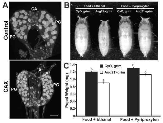
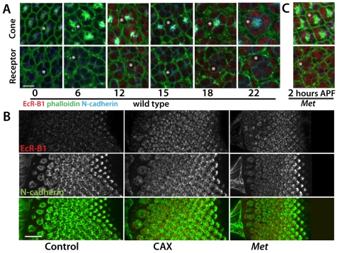
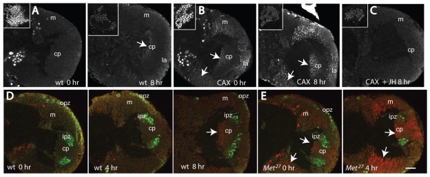
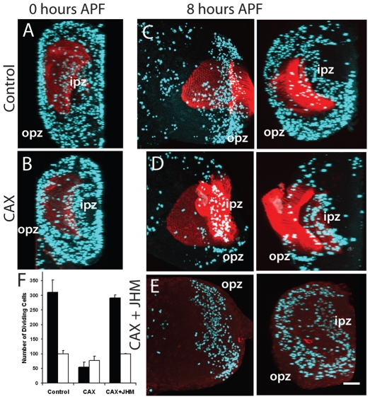
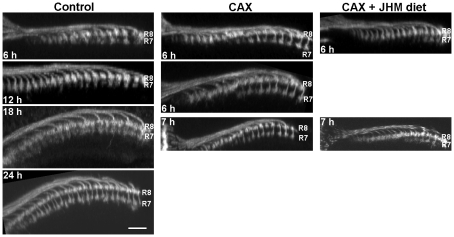
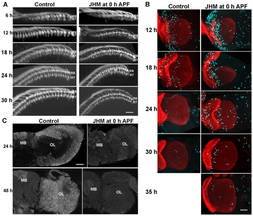
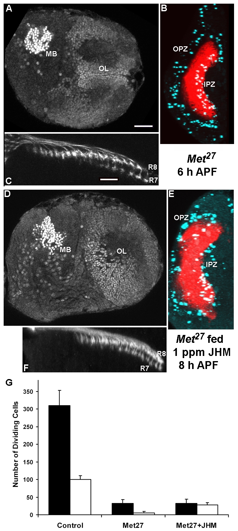
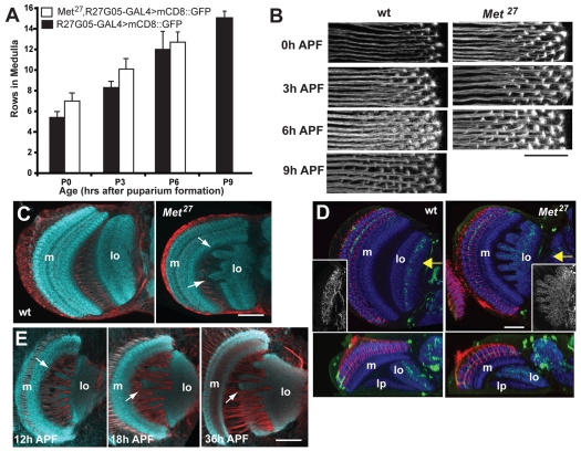
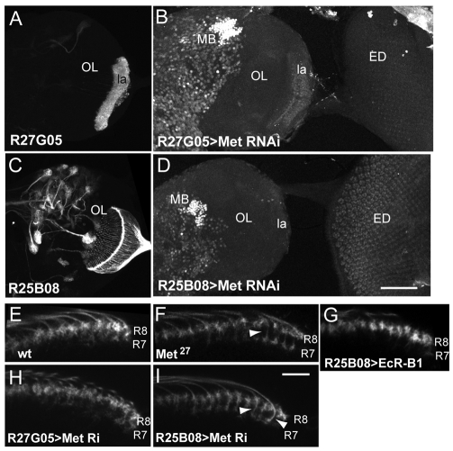
To elucidate the role of juvenile hormone (JH) in metamorphosis of Drosophila melanogaster, the corpora allata cells, which produce JH, were killed using the cell death gene grim. These allatectomized (CAX) larvae were smaller at pupariation and died at head eversion. They showed premature ecdysone receptor B1 (EcR-B1) in the photoreceptors and in the optic lobe, downregulation of proliferation in the optic lobe, and separation of R7 from R8 in the medulla during the prepupal period. All of these effects of allatectomy were reversed by feeding third instar larvae on a diet containing the JH mimic (JHM) pyriproxifen or by application of JH III or JHM at the onset of wandering. Eye and optic lobe development in the Methoprene-tolerant (Met)-null mutant mimicked that of CAX prepupae, but the mutant formed viable adults, which had marked abnormalities in the organization of their optic lobe neuropils. Feeding Met(27) larvae on the JHM diet did not rescue the premature EcR-B1 expression or the downregulation of proliferation but did partially rescue the premature separation of R7, suggesting that other pathways besides Met might be involved in mediating the response to JH. Selective expression of Met RNAi in the photoreceptors caused their premature expression of EcR-B1 and the separation of R7 and R8, but driving Met RNAi in lamina neurons led only to the precocious appearance of EcR-B1 in the lamina. Thus, the lack of JH and its receptor Met causes a heterochronic shift in the development of the visual system that is likely to result from some cells 'misinterpreting' the ecdysteroid peaks that drive metamorphosis.


Figures

Fig. 1.
Fig. 1.
Genetic allatectomy and its effect on puparial size. (A)The ring gland of control CyO, UAS-grim (above) and allatectomized (CAX) Aug21>grim (below) wandering larvae showing the loss of the corpora allata (CA) in the latter. a, aorta; PG, prothoracic gland. Scale bar: 25 μm. (B,C) Sample puparia (B) and the pupal weights (C) of CyO, UAS-grim (control) and Aug21>grim (allatectomized) larvae raised on normal diet containing 1% ethanol or 1 ppm pyriproxyfen (JHM) in 1% ethanol. Bars represent the average ± s.d. for 50-60 puparia (one day after wandering). Different letters denote average weights that are significantly different (P≤0.0001, using one-way ANOVA and Tukey-Kramer HSD comparison of means).
Fig. 2.
Fig. 2.
Developmental appearance of the ecdysone receptor EcR-B1 in the photoreceptors in CyO, UAS-grim (control), Aug21>grim (allatectomized, CAX), and Met27 prepupae and pupae. (A) Confocal sections of developing ommatidia at the levels of the crystalline cone cells (top) and the photoreceptors (bottom) showing the time course of appearance of EcR-B1 (red) in wild-type prepupae and pupae. Cellular outlines are delineated by phalloidin staining (green). Times shown are hours after puparium formation (APF). Note that EcR-B1 is first evident at 12 hours APF. Scale bar: 5 μm. (B) Confocal sections showing the precocious presence of EcR-B1 in the photoreceptors of allatectomized (CAX) and Met27 eye discs at the time of pupariation. N-cadherin (green) stains developing photoreceptors. Scale bar: 20 μm. (C) Ommatidia from the eye disc of a Met27 prepupa at 2 hours APF showing the precocious appearance of EcR-B1. In A and C, the asterisks indicate an example of a cone cell (top) or a photoreceptor (bottom).
Fig. 3.
Fig. 3.
Precocious appearance of the ecdysone receptor EcR-B1 in the optic lobes (OL) of Aug21>grim allatectomized (CAX) prepupae and its suppression by feeding larvae on a diet containing 1 ppm pyriproxyfen (JHM). (A-E) OL confocal sections at different times (X hours; hr) APF of (A,D) CyO, UAS-grim (control) prepupae, (B) CAX prepupae after feeding on normal diet, (C) CAX prepupa after feeding on the JHM diet, and (E) Met27 prepupae. Arrows indicate EcR-B1-positive neurons. Insets in A-C show endogenous EcR-B1 in the mushroom body neurons. (D,E) Deadpan-positive neuroblasts (green); EcR-B1 neurons (red). cp, ‘central plug’ neurons produced by the neuroblasts of the inner proliferation zone (ipz); la, lamina; m, medulla; opz, outer proliferation zone. Scale bar: 25 μm.
Fig. 4.
Fig. 4.
Precocious decline in proliferation in the OL in Aug21>grim allatectomized (CAX) prepupae and its prevention by feeding larvae on diet containing 1 ppm pyriproxyfen (JHM). (A-E) The OL at different times (hours) APF of (A,C) CyO, UAS-grim (control) prepupae, (B,D) CAX prepupae after feeding on normal diet, and (E) a CAX prepupa after feeding on the JHM diet. (C-E) Frontal z-projection of the optic lobe (left) and a 90° projection along the x-axis (right), showing an end-on view to discriminate the inner (ipz) and outer (opz) proliferation zones; (A,B) x-axis projections. Aqua represents PH3 staining of mitotic cells; red is chaoptin staining of the incoming photoreceptors, except in E where it was omitted. Scale bar: 50 μm. (F) Mean number (±s.d.) of proliferating cells in the opz (black bars) and the ipz (white bars) of 3-5 animals at 8 hours APF.
Fig. 5.
Fig. 5.
Dorsal views of the medulla showing the effects of JH manipulations on the timing of separation of the R7 and R8 growth cones. (Left) Control prepupae showing that growth cones become completely separated by 18 to 24 hours (h) APF. (Middle) Allatectomized (CAX) prepupae show complete separation by 6 hours APF. (Right) Feeding CAX larvae on a JHM diet blocks the precocious separation. Scale bar: 10 μm.
Fig. 6.
Fig. 6.
The effect of 100 ng pyriproxifen (JHM) applied at the time of pupariation on OL development. (A) Dorsal view of the medulla showing that JHM treatment retards the separation of the R7 and R8 growth cones. (B) Frontal projections of the optic lobe showing the prolongation of proliferation in the JHM-treated animals: red, chaoptin; aqua, phosphohistone H3. (C) JHM treatment suppresses the appearance of EcR-B1. h, hours APF; MB, mushroom body. Scale bars: 10 μm in A; 25 μm in B,C.
Fig. 7.
Fig. 7.
The Met27 mutant shows precocious OL development and reduced sensitivity to a JHM (1 ppm pyriproxifen) diet. (A,D) Frontal sections through the brain and OL showing precocious EcR-B1 immunostaining in the OL of both Met27 prepupae (A) and those fed a JHM diet (D). (B,E) An x-projection of the OL showing that activity in the outer proliferation zone (OPZ) is also precociously reduced in both groups. red, chaoptin; aqua, phosphohistone H3; IPZ, inner proliferation zone. (C,F) Dorsal view of the medulla showing that the prominent separation of the R7 and R8 growth cones is partially suppressed by feeding the mutant JHM. In the above series, the Met27 examples were at 6 hours APF and the Met27 fed JHM were at 8 hours APF. (G) Mean number (±s.d.) of proliferating cells in the OPZ (black bars) and IPZ (white bars) of 4-5 animals at 8 hours APF. The reduced proliferation in the OPZ is not rescued in the mutants that were fed on the JHM diet, whereas the number of dividing cells in the IPZ is partially restored. Scale bars: in A, 25 μm for A,B,D,E; in C, 10 μm for C,F.
Fig. 8.
Fig. 8.
Comparison of OL development in wild-type and Met27 mutants. (A,B) R27G05-GAL4 driving mCD8::GFP revealed lamina axon ingrowth into the medulla in wild-type and Met27 backgrounds. (A) The mean number (±s.d.) of rows of lamina neuron terminals seen in the medulla of wild-type (black bars) and Met27 (white) individuals at various times after pupariation; 3-5 samples per bar. (B) Confocal projections of rows of lamina neuron terminals in the medulla at various times APF. Scale bar: 10 μm. (C,D) Frontal sections of the adult OL. (C) The lobula (lo) of the Met27 mutant has abnormal lobes (arrows) that project towards the medulla (m); (D) expression of GFP in the R23C06 line reveals lobula interneurons whose dendrites are normally confined to defined layers of the lobula, but in the mutant they extend throughout this neuropil: red, chaoptin; blue, N-cadherin. Insets show a z-projections of the entire dendritic tree. Below are horizontal sections at the level of the arrow; lp, lobula plate. (E) Sections through the developing optic lobe of Met27 mutants show that lobula irregularities are evident as early as 12 hours APF. Staining in C and E: N-cadherin, aqua; neuroglian, red. Scale bars: 20 μm in B; 50 μm in C-E.
Fig. 9.
Fig. 9.
Effects of targeted expression of genes involved in the response to JH. (A,C) Images of wandering larval brains showing that (A) R27G05 drives expression in lamina neurons (la), and (C) R25B08 drives expression in the photoreceptors, but not in any intrinsic neurons in the lamina or medulla. (B,D) Confocal projection showing EcR-B1 expression in the brain hemisphere and attached eye imaginal disc (ED) at 3 hours APF. Expression of Met RNAi under the control of R27G05 and R25B08 results in precocious appearance of EcR-B1 in lamina neurons (la) and photoreceptors, respectively. Mushroom bodies (MB) show strong endogenous expression. (E-I) Transverse slices through the medulla at 6 hours APF, showing the relative position of the terminals of photoreceptors R7 and R8. (E) Control showing the overlap of R7 and R8. F) Met27 mutant in which R7 appears to have a distinct stalk (arrowhead) that separates it from R8. (G) Expression of EcR-B1 in the photoreceptors does not produce an early separation of the two terminals. (H) Knockdown of Met in lamina neurons by RNAi does not produce a separation. (I) Knockdown of Met in the photoreceptors results in early, stalked (arrowheads) terminals for R7. Scale bars: in D, 100 μm for A-D; in I, 10 μm for E-I.

Similar articles

Cited by

References

    1. Ashok M., Turner C., Wilson T. G. (1998). Insect juvenile hormone resistance gene homology with the bHLH-PAS family of transcriptional regulators. Proc. Natl. Acad. Sci. USA 95, 2761-2766 - PMC - PubMed
    1. Boone J. Q., Doe C. Q. (2008). Identification of Drosophila Type II neuroblast lineages containing transit amplifying ganglion mother cells. Dev. Neurobiol. 68, 1185-1195 - PMC - PubMed
    1. Bownes M., Rembold H. (1987). The titre of juvenile hormone during the pupal and adult stages of the life cycle of Drosophila melanogaster. Eur. J. Biochem. 164, 709-712 - PubMed
    1. Champlin D. T., Truman J. W. (1998a). Ecdysteroids govern two phases of eye development during metamorphosis of the moth, Manduca sexta. Development 125, 2009-2018 - PubMed
    1. Champlin D. T., Truman J. W. (1998b). Ecdysteroid control of cell proliferation during optic lobe neurogenesis in the moth, Manduca sexta. Development 125, 269-277 - PubMed

Publication types

MeSH terms

LinkOut - more resources