. 2010 Apr;137(7):1159-67.
doi: 10.1242/dev.045005. Epub 2010 Feb 24.

Runx1 and Runx2 cooperate during sternal morphogenesis

Affiliations

Runx1 and Runx2 cooperate during sternal morphogenesis

Ayako Kimura et al. Development. 2010 Apr.

Abstract

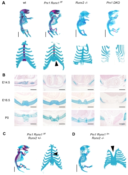
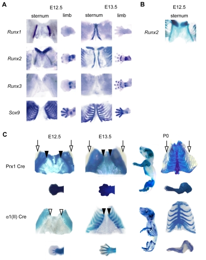
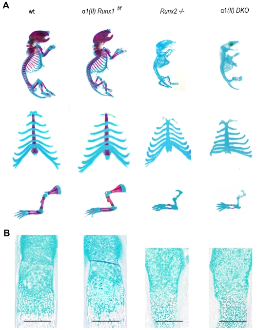
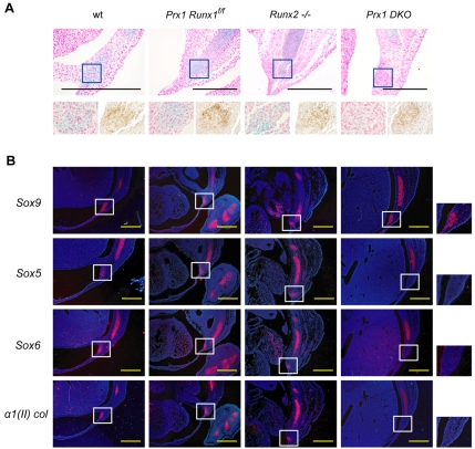
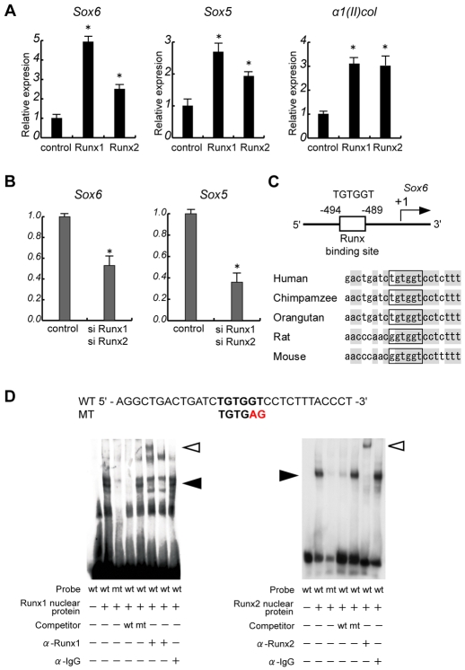
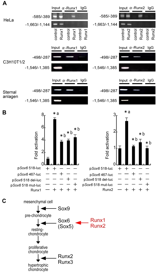
Chondrocyte differentiation is strictly regulated by various transcription factors, including Runx2 and Runx3; however, the physiological role of Runx1 in chondrocyte differentiation remains unknown. To examine the role of Runx1, we generated mesenchymal-cell-specific and chondrocyte-specific Runx1-deficient mice [Prx1 Runx1(f/f) mice and alpha1(II) Runx1(f/f) mice, respectively] to circumvent the embryonic lethality of Runx1-deficient mice. We then mated these mice with Runx2 mutant mice to obtain mesenchymal-cell-specific or chondrocyte-specific Runx1; Runx2 double-mutant mice [Prx1 DKO mice and alpha1(II) DKO mice, respectively]. Prx1 Runx1(f/f) mice displayed a delay in sternal development and Prx1 DKO mice completely lacked a sternum. By contrast, alpha1(II) Runx1(f/f) mice and alpha1(II) DKO mice did not show any abnormal sternal morphogenesis or chondrocyte differentiation. Notably, Runx1, Runx2 and the Prx1-Cre transgene were co-expressed specifically in the sternum, which explains the observation that the abnormalities were limited to the sternum. Histologically, mesenchymal cells condensed normally in the prospective sternum of Prx1 DKO mice; however, commitment to the chondrocyte lineage, which follows mesenchymal condensation, was significantly impaired. In situ hybridization analyses demonstrated that the expression of alpha1(II) collagen (Col2a1 - Mouse Genome Informatics), Sox5 and Sox6 in the prospective sternum of Prx1 DKO mice was severely attenuated, whereas Sox9 expression was unchanged. Molecular analyses revealed that Runx1 and Runx2 induce the expression of Sox5 and Sox6, which leads to the induction of alpha1(II) collagen expression via the direct regulation of promoter activity. Collectively, these results show that Runx1 and Runx2 cooperatively regulate sternal morphogenesis and the commitment of mesenchymal cells to become chondrocytes through the induction of Sox5 and Sox6.


Figures

Fig. 1.
Fig. 1.
Sternal abnormality in mesenchymal-specific Runx1-deficient mice. (A,C,D) Skeletal preparation of newborn mice. (A) Note the delay in calcification of the xiphoid process in Prx1 Runx1f/f mice (arrowhead) and the absence of the sternum in Prx1 DKO mice. By contrast, the sternal bars of Runx2−/− mice are fused by birth, yet their xiphoid processes are non-mineralized. (A,C) Prx1 Runx1f/f/Runx2+/− mice show an intermediate phenotype between Prx1 Runx1f/f mice and Prx1 DKO mice (arrowhead). (A,D) Prx1 Runx1f/+/Runx2−/− mice show an intermediate phenotype between Runx2−/− mice and Prx1 DKO mice (arrowhead). (B) Histological analysis of mouse embryo sternums. Axial section. Note the absence of the sternum and the protrusion of internal organs in Prx1 DKO mice. Prx1 Runx1f/f mice and Prx1 DKO mice denote Prx1 Cre; Runx1f/f mice and Prx1 Cre; Runx1f/f; Runx2−/− mice, respectively. Scale bars: 5 mm in A,C,D; 500 μm in B.
Fig. 2.
Fig. 2.
Expression of the Runx genes, the Prx1-Cre transgene and the α1(II) collagen-Cre transgene during skeletal development. (A) Whole-mount in situ hybridization analysis of the Runx genes and Sox9 in E12.5 and 13.5 mouse embryos. Runx1 and Runx2, but not Runx3, are expressed in the prospective sternum. (B) lacZ staining analysis of Runx2+/− mice. Note the distinct expression of Runx2 as shown by positive blue staining in the prospective sternum. (C) lacZ staining of Prx1-Cre; Rosa26 reporter and α1(II)-Cre; Rosa26 reporter embryos. Note that the Prx1-Cre transgene is expressed as early as E12.5 in the prospective sternum, whereas the α1(II)-Cre transgene is expressed only after E13.5. Expression of the Prx1-Cre transgene was also observed in the ventral, but not dorsal (arrows), rib cage from E12.5 to birth (P0). Arrowheads indicate the future sternum where the Cre transgenes are (black) or are not (white) expressed.
Fig. 3.
Fig. 3.
Normal chondrocyte differentiation in committed chondrocyte-specific Runx1-deficient mice. (A) Skeletal preparations of newborn mice. (B) Histological sections of femurs from newborn mice. Sections were stained with Alcian Blue and Nuclear Fast Red stain; extracellular cartilage matrix is stained blue. There are no overt differences between wild-type and α1(II)-Cre; Runx1f/f mice or between Runx2−/− mice and α1(II)-Cre DKO mice. α1(II) Runx1f/f mice and α1(II) DKO mice denote α1(II)-Cre; Runx1f/f mice and α1(II)-Cre; Runx1f/f; Runx2−/− mice, respectively. Scale bars: 500 μm.
Fig. 4.
Fig. 4.
Normal condensation but abolished expression of Sox5 and Sox6 in the prospective sternum of Prx1-Cre DKO mice. (A) Histological analysis of axial sections of E13.5 mouse embryo prospective sternums. Upper and bottom left, Alcian Blue staining; bottom right, PNA staining. The bottom figures show high magnification of each corresponding rectangle in the upper panel. Note the PNA-positive mesenchymal condensations in Prx1 DKO mice. The diminished Alcian Blue staining in Prx1 DKO mice suggests the impaired accumulation of cartilage matrices. (B) In situ hybridization analysis of E13.5 mouse embryo prospective sternums (boxed area). In Prx1-Cre DKO mice, Sox9 expression in the prospective sternum (white box and higher magnification in the right panel) is not altered, whereas Sox5, Sox6 and α1(II) collagen expression in the same area are markedly decreased. Prx1 Runx1f/f mice and Prx1 DKO mice denote Prx1-Cre; Runx1f/f mice and Prx1-Cre; Runx1f/f; Runx2−/− mice, respectively. Scale bars: 500 μm.
Fig. 5.
Fig. 5.
Sox6 is a molecular target of Runx1 and Runx2 in chondrocyte differentiation. (A,B) Real-time PCR analysis. (A) Runx1 and Runx2 induced the expression of Sox6 (left), Sox5 (middle) and α1(II) collagen (right) in C3H10T1/2 cells. (B) The knockdown of Runx1 and Runx2 significantly decreased the expression of Sox6 and Sox5 in C3H10T1/2 cells. (C) Schematic representation of the putative Runx-binding site in the Sox6 promoter (top). Comparison of the sequence of the putative Runx-binding site in the Sox6 promoter across different species (bottom). Note the conservation of the binding site across species. (D) EMSA. Nuclear extracts (NEs) from Runx1- (left) or Runx2- (right) expressing COS cells formed a protein-DNA complex following incubation with oligonucleotides encompassing the Runx binding site in the Sox6 promoter (black arrowheads). NEs incubated with antibodies against Runx1 or Runx2 showed supershifted bands (white arrowheads). *, P<0.05 to control.
Fig. 6.
Fig. 6.
Sox6 is a molecular target of Runx1 and Runx2 in chondrocyte differentiation. (A) ChIP assay. An antibody against Runx1 (left) or Runx2 (right) immunoprecipitated the Runx binding site of the human Sox6 promoter (−494/−489) in HeLa cells overexpressing Runx1 and Runx2 (top). The same antibody also immunoprecipitated Runx binding site of the mouse Sox6 promoter (−454/−449) in untransfected mesenchymal-chondrogenic C3H10T1/2 cells (middle) and naive mouse primary sternal anlagen (bottom). (B) Sox6 promoter activity in HeLa cells. Runx1 (left) or Runx2 (right) significantly increased the activity of the 0.5 kb Sox6 promoter (pSox6 518-luc). This induction was reduced by 50% by deleting (pSox6 467-luc, pSox6 518 del-luc) or mutating (pSox6 518 mut-luc) the Runx binding site in the promoter. *a: P<0.05 to control. *b: P<0.05 to pSox6 518-luc overexpressing Runx protein. (C) Proposed mechanism: Runx1 and Runx2 cooperatively upregulate Sox5 and Sox6 expression, which in turn induces chondrocyte differentiation.

Similar articles

Cited by

References

    1. Akiyama H., Chaboissier M., Martin J., Schedl A., de Crombrugghe B. (2002). The transcription factor Sox9 has essential roles in successive steps of the chondrocyte differentiation pathway and is required for expression of Sox5 and Sox6. Genes Dev. 16, 2813-2828 - PMC - PubMed
    1. Bi W., Deng J. M., Zhang Z., Behringer R. R., de Crombrugghe B. (1999). Sox9 is required for cartilage formation. Nat. Genet. 22, 85-89 - PubMed
    1. Chen J. (1952). Studies on the morphogenesis of the mouse sternum. II. Experiments on the origin of the sternum and its capacity for self-differentiation in vitro. J. Anat. 86, 387-401 - PMC - PubMed
    1. Day T., Guo X., Garrett-Beal L., Yang Y. (2005). Wnt/beta-catenin signaling in mesenchymal progenitors controls osteoblast and chondrocyte differentiation during vertebrate skeletogenesis. Dev. Cell 8, 739-750 - PubMed
    1. de Crombrugghe B., Lefebvre V., Nakashima K. (2001). Regulatory mechanisms in the pathways of cartilage and bone formation. Curr. Opin. Cell Biol. 13, 721-727 - PubMed

Publication types

MeSH terms