

Transforming growth factor-{beta} coordinately induces suppressor of cytokine signaling 3 and leukemia inhibitory factor to suppress osteoclast apoptosis
- PMID: 20181800
- PMCID: PMC2850239
- DOI: 10.1210/en.2009-0813
Transforming growth factor-{beta} coordinately induces suppressor of cytokine signaling 3 and leukemia inhibitory factor to suppress osteoclast apoptosis
Abstract
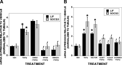
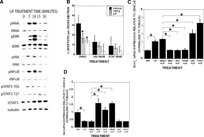
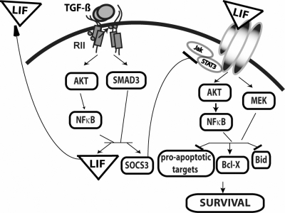
Local release of TGF-beta during times of high bone turnover leads to elevated levels within the bone microenvironment, and we have shown that TGF-beta suppresses osteoclast apoptosis. Therefore, understanding the influences of TGF-beta on bone resorbing osteoclasts is critical to the design of therapies to reduce excess bone loss. Here we investigated the mechanisms by which TGF-beta sustains suppression of osteoclast apoptosis. We found TGF-beta rapidly increased leukemia inhibitory factor (LIF) expression and secretion by phosphorylated mothers against decapentaplegic-dependent and -independent signaling pathways. TGF-beta also induced suppressor of cytokine signaling 3 (SOCS3) expression, which was required for TGF-beta or LIF to promote osteoclast survival by. Blocking LIF or SOCS3 blocked TGF-beta promotion of osteoclast survival, confirming that LIF and SOCS3 expression are necessary for TGF-beta-mediated suppression of osteoclast apoptosis. Investigation of the mechanisms by which LIF promotes osteoclast survival revealed that LIF-induced expression of Bcl-X(L) and repressed Bcl-2 interacting domain expression by activating MAPK kinase, AKT, and nuclear factor-kappaB pathways. Suppression of Janus kinase/signal transducer and activator of transcription signaling further increased Bcl-X(L) expression and enhanced osteoclast survival, supporting that this pathway is not involved in prosurvival effects of TGF-beta and LIF. These data show that TGF-beta coordinately induces LIF and SOCS3 to promote prosurvival signaling. This alters the ratio of prosurvival Bcl2 family member Bcl-X(L) to proapoptotic family member Bcl-2 interacting domain, leading to prolonged osteoclast survival.
Figures
Similar articles
-
TGF-beta coordinately activates TAK1/MEK/AKT/NFkB and SMAD pathways to promote osteoclast survival.Exp Cell Res. 2008 Sep 10;314(15):2725-38. doi: 10.1016/j.yexcr.2008.06.006. Epub 2008 Jun 13. Exp Cell Res. 2008. PMID: 18586026 Free PMC article.
-
Leukemia inhibitory factor induces an antiapoptotic response in oligodendrocytes through Akt-phosphorylation and up-regulation of 14-3-3.Proteomics. 2008 Mar;8(6):1237-47. doi: 10.1002/pmic.200700641. Proteomics. 2008. PMID: 18338825
-
The effect of FK506 on transforming growth factor beta signaling and apoptosis in chronic lymphocytic leukemia B cells.Haematologica. 2008 Jul;93(7):1039-48. doi: 10.3324/haematol.12402. Epub 2008 May 19. Haematologica. 2008. PMID: 18492692
-
Signaling Cross Talk between TGF-β/Smad and Other Signaling Pathways.Cold Spring Harb Perspect Biol. 2017 Jan 3;9(1):a022137. doi: 10.1101/cshperspect.a022137. Cold Spring Harb Perspect Biol. 2017. PMID: 27836834 Free PMC article. Review.
-
TGF-beta signaling and the role of inhibitory Smads in non-small cell lung cancer.J Thorac Oncol. 2010 Apr;5(4):417-9. doi: 10.1097/JTO.0b013e3181ce3afd. J Thorac Oncol. 2010. PMID: 20107423 Free PMC article. Review.
Cited by
-
Stimulation of osteoclast migration and bone resorption by C-C chemokine ligands 19 and 21.Exp Mol Med. 2017 Jul 21;49(7):e358. doi: 10.1038/emm.2017.100. Exp Mol Med. 2017. PMID: 28729639 Free PMC article.
-
Therapeutic and Preventive Effects of Osteoclastogenesis Inhibitory Factor on Osteolysis, Proliferation of Mammary Tumor Cell and Induction of Cancer Stem Cells in the Bone Microenvironment.Int J Mol Sci. 2018 Mar 16;19(3):888. doi: 10.3390/ijms19030888. Int J Mol Sci. 2018. PMID: 29547583 Free PMC article.
-
Non-Coding RNAs in Multiple Myeloma Bone Disease Pathophysiology.Noncoding RNA. 2020 Sep 9;6(3):37. doi: 10.3390/ncrna6030037. Noncoding RNA. 2020. PMID: 32916806 Free PMC article. Review.
-
New insights into Chlamydia pathogenesis: Role of leukemia inhibitory factor.Front Cell Infect Microbiol. 2022 Oct 18;12:1029178. doi: 10.3389/fcimb.2022.1029178. eCollection 2022. Front Cell Infect Microbiol. 2022. PMID: 36329823 Free PMC article. Review.
-
Glucagon-like peptide 2 decreases osteoclasts by stimulating apoptosis dependent on nitric oxide synthase.Cell Prolif. 2018 Aug;51(4):e12443. doi: 10.1111/cpr.12443. Epub 2018 Feb 19. Cell Prolif. 2018. PMID: 29457300 Free PMC article.
References
-
- Guise TA, Mundy GR 1998 Cancer and bone. Endocr Rev 19:18–54 - PubMed
-
- Michigami T, Ihara-Watanabe M, Yamazaki M, Ozono K 2001 Receptor activator of nuclear factor κB ligand (RANKL) is a key molecule of osteoclast formation for bone metastasis in a newly developed model of human neuroblastoma. Cancer Res 61:1637–1644 - PubMed
-
- Roodman GD 1995 Osteoclast function in Paget’s disease and multiple myeloma. Bone 17:57S–61S - PubMed
-
- Winding B, Misander H, Sveigaard C, Therkildsen B, Jakobsen M, Overgaard T, Oursler MJ, Foged NT 2000 Human breast cancer cells induced angiogenesis, recruitment, and activation of osteoclasts in osteolytic metastasis. J Cancer Res Clin Oncol 126:631–640 - PubMed
Publication types
MeSH terms
Substances
Grants and funding
LinkOut - more resources
Full Text Sources
Molecular Biology Databases
Research Materials
