. 2010 Apr 15;21(8):1362-74.
doi: 10.1091/mbc.e09-07-0546. Epub 2010 Feb 24.

Mso1p regulates membrane fusion through interactions with the putative N-peptide-binding area in Sec1p domain 1

Affiliations

Mso1p regulates membrane fusion through interactions with the putative N-peptide-binding area in Sec1p domain 1

Marion Weber et al. Mol Biol Cell. .

Abstract

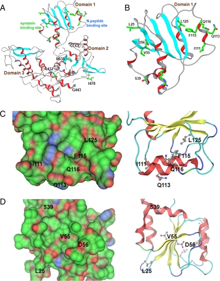
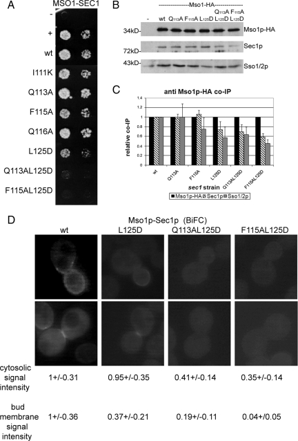
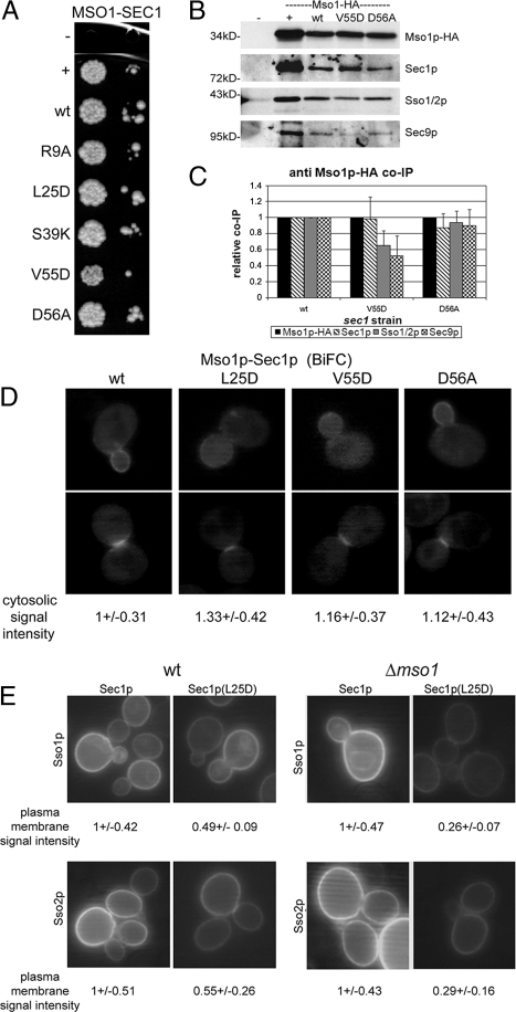
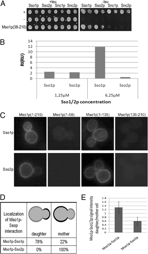
Sec1p/Munc18 (SM) family proteins regulate SNARE complex function in membrane fusion through their interactions with syntaxins. In addition to syntaxins, only a few SM protein interacting proteins are known and typically, their binding modes with SM proteins are poorly characterized. We previously identified Mso1p as a Sec1p-binding protein and showed that it is involved in membrane fusion regulation. Here we demonstrate that Mso1p and Sec1p interact at sites of exocytosis and that the Mso1p-Sec1p interaction site depends on a functional Rab GTPase Sec4p and its GEF Sec2p. Random and targeted mutagenesis of Sec1p, followed by analysis of protein interactions, indicates that Mso1p interacts with Sec1p domain 1 and that this interaction is important for membrane fusion. In many SM family proteins, domain 1 binds to a N-terminal peptide of a syntaxin family protein. The Sec1p-interacting syntaxins Sso1p and Sso2p lack the N-terminal peptide. We show that the putative N-peptide binding area in Sec1p domain 1 is important for Mso1p binding, and that Mso1p can interact with Sso1p and Sso2p. Our results suggest that Mso1p mimics N-peptide binding to facilitate membrane fusion.


Figures

Figure 1.
Figure 1.
Localization of the Mso1p–Sec1p interaction site in vivo. (A) Live cell imaging of vegetatively grown haploid (H304) cells expressing YFP(C)-Mso1p (B3044) or YFP(C)-Mso1p(136-210) (B3064) with Sec1p-Venus(N) (B2930). (B) Live cell imaging of vegetatively grown haploid cells (H304) expressing YFP(C)-Mso1p(1-58) (B3355), YFP(C)-Mso1p(1-135) (B3354), YFP(C)-Mso1p(1-210) (B3044), YFP(C)-Mso1p(59-210) (B3353), or YFP(C)-Mso1p(136-210) (B3064) with Sec1p-Venus(N) (B2930), and (C) diploid (H2530) cells expressing Mso1p-Venus(C) (B2918) or Mso1p(136-210)-Venus(C) (B3012) with Sec1p-Venus(N) (B2930). Mso1p–Sec1p interaction is detected preferentially at the bud tip, plasma membrane of the growing bud, and the bud neck (arrowheads). Regularly, the interaction signal appeared dott-like (arrows). The interaction site was adjacent to the bud or at the opposite pole of the cells (star). (D) Mso1p–Sec1p interaction is altered in different secretion mutants. Indicated mutants cells expressing YFP(C)-Mso1p (B3044) with Sec1p-Venus(N) (B2930) were grown to OD600 0.8–1 at 24°C, split, and either left at 24°C or shifted to 37°C for 1 h before investigation. Mislocalization and clusters of the interaction signal are pointed out by arrows or a dotted line.
Figure 2.
Figure 2.
Mso1p stability is depended on Sec1p. (A) The wild-type, sec1-1, sec1-11, sec2-41 and sec4-8 strains where the sole copy of MSO1 is HA tagged were grown to an OD600 of 1 at 24°C, split, and either maintained at 24°C or shifted to 37°C for 1 h before lysate preparation. Lysates were subjected to Western blotting and detection with anti-HA and anti-Sso1p/2p antibodies. (B) Overexpression of SEC1 restores Mso1p levels. A SEC1 overexpression vector or an empty vector was transformed to wild-type and sec1-1 and sec1-11 cells expressing Mso1p-HA. The temperature shift experiment was performed as is in (A). Detection was done with anti-HA and anti-Sec1p antibodies. (C) Sec1p level is not affected by MSO1 deletion. HA-tagged Sec1p was detected in wild-type and Δmso1 cells. (A–C) The quantification of the ECL signals, normalized against the wild-type 24°C lysate, are shown underneath each lane.
Figure 3.
Figure 3.
Mapping of the Mso1p-binding site in Sec1p. (A) Map of the Mso1p interacting (blue), weakly interacting (light blue), and noninteracting (white) sec1 mutations. The insertion sites are indicated by black vertical lines within the SEC1 gene. Twenty mutants were sequenced to confirm the mapping. The domain borders of Sec1p are indicated above of the gene. (B) Yeast two-hybrid assay between different Mso1p fragments and Sec1p domains 1 and 3. Mso1p (59-210) is lacking the Sec1p-binding area (Knop et al., 2005). As an additional negative control, Bicoid (B1228) and as a positive control Gal4 (B1229) were used. Two 10-fold dilutions of cells were grown with or without leucine.
Figure 4.
Figure 4.
A model for Sec1p structure. (A) The overall structure of Sec1p. Two potential protein-binding interfaces can be identified: the putative syntaxin binding and the N-peptide–binding site. (B) A detailed view of domain 1 where the potential N-peptide and syntaxin-like binding sites are located. Helices are depicted in red and strands in light blue. Only side chains of residues that were subjected to site targeted mutagenesis are shown. (C and D) Surface and ribbon presentations of the putative N-peptide– (C) and syntaxin- (D) binding sites. The Connolly surface is colored green for hydrophobic, red for hydrogen-bonding, and blue for mildly polar atoms. Only positions of residues mutated in the present study are indicated.
Figure 5.
Figure 5.
Mso1p binding is affected by mutations in the putative N-peptide binding area in Sec1p. (A) Yeast two-hybrid assay between Mso1p and different Sec1p mutants. As a negative control Bicoid (B1228) and as a positive control Gal4 (B1229) were used. Two 10-fold dilutions of cells grown on medium without leucine are shown. (B and C) Mso1p coimmunoprecipitates less efficiently with Sec1p N-peptide domain mutants. Cells with endogenously HA-tagged Mso1p expressing different sec1 mutants were grown to OD600 = 1, lysed, and subjected to anti-HA immunoprecipitations. Immunoprecipitates were subjected to Western blotting and detection with anti-HA, Sec1p, and Sso1p/2p antibodies. (B) A representative Western of the immunoprecipitations. (C) Quantification of independent immunoprecipitation normalized to the amount of immunoprecipitated Mso1p-HA. SDs are shown for each immunoprecipitation. (D) Localization of the Mso1p interaction site with Sec1p mutants L125D, Q113A L125D, and F115 L125D. Haploid vegetatively grown cells (H304) expressing YFP(C)-Mso1p (B3044) and different mutant versions of Sec1p-Venus(N) [wt (B2930), L125D (B3263), Q113A L125D (B3264), and F115A L125D (B3265)] were investigated by fluorescence microscopy. The BiFC signal was quantified as described in Materials and Methods.
Figure 6.
Figure 6.
SNARE complex, but not Mso1p binding is affected in sec1 (L25D) and sec1 (V55D) mutants. (A) Yeast two-hybrid assay between Mso1p and different Sec1p syntaxin-binding mutants. As a negative control Bicoid (B1228) and as a positive control Gal4 (B1229) was used. The interactions were scored of two 10-fold dilutions on medium lacking leucine. In B and C less coimmunoprecipitation of Sso1p/2p and Sec9p is observed in Sec1p- (V55D) and Sec1p- (D56A) expressing cells. MSO1-HA cells expressing different sec1 mutants were grown until a OD600 = 1, lysed, and subjected to anti-HA immunoprecipitations. Immunoprecipitates were analyzed by Western blotting, and the blots detected with anti-HA, Sec1p, Sso1p/2p, and Sec9 antibodies. (B) A representative Western blot of the immunoprecipitations. (C) Quantification of independent immunoprecipitations, normalized to the amount of immunoprecipitated Mso1p-HA. SDs are shown for each immunoprecipitation. (D) Localization of the Mso1p interaction site with Sec1p mutants L25D, V55D, and D56A in vivo. Haploid, vegetatively grown cells (H304) expressing YFP(C)-Mso1p (B3044) and different mutant versions of Sec1p-Venus(N) [wt (B2930), L25D (B3260), V55D (B3261), and D56A (B3262)] were investigated by fluorescence microscopy. (E) Sec1p(L25D) BiFC interaction with Sso1p and Sso2p is sensitive to deletion of MSO1. Haploid vegetatively grown cells [wt (H304) or Δmso1 (H2658)] expressing Sec1p-YFP(C) wt (B3308) or the L25D (B3312) mutant together with either YFP(N)-Sso1p (B3307) or YFP(N)-Sso2p (B3309) were investigated by fluorescence microscopy for YFP signal. The BiFC signal was quantified as described in Materials and Methods..
Figure 7.
Figure 7.
Mso1p interacts with Sso1p and Sso2p. (A) Yeast two-hybrid assay between Mso1p and Sso1p, Sso2p, Snc1p, and Snc2p. As negative control Bicoid (B1228) and as positive control Gal4 (B1229) was used. Two 10-fold dilutions on medium with or without leucine are shown. (B) In vitro analysis of Sso protein interaction with Mso1p. The interactions were analyzed by the surface plasmon resonance technique as described in Materials and Methods. (C) Mso1p interacts with Sso1p/2p at the plasma membrane. Haploid vegetatively grown cells expressing YFP(C)-Mso1p (B3044), YFP(C)-Mso1p(1-58) (B3355), YFP(C)-Mso1p(1-135) (B3354), YFP(C)-Mso1p(136-210) (B3043), and YFP(N)-Sso1p/2p (B3307, B3309) were investigated by fluorescence microscopy. (D) Quantification of the Mso1p–Ssop localization. (E) Quantification of the Mso1p-Ssop plasma membrane signal intensity in daughter versus mother cell.

Similar articles

Cited by

References

    1. Aalto M. K., Jantti J., Ostling J., Keranen S., Ronne H. Mso1p: a yeast protein that functions in secretion and interacts physically and genetically with Sec1p. Proc. Natl. Acad. Sci. USA. 1997;94:7331–7336. - PMC - PubMed
    1. Biederer T., Sudhof T. C. Mints as adaptors: direct binding to neurexins and recruitment of munc18. J. Biol. Chem. 2000;275:39803–39806. - PubMed
    1. Bracher A., Weissenhorn W. Crystal structures of neuronal squid Sec1 implicate inter-domain hinge movement in the release of t-SNAREs. J. Mol. Biol. 2001;306:7–13. - PubMed
    1. Bracher A., Weissenhorn W. Structural basis for the Golgi membrane recruitment of Sly1p by Sed5p. EMBO J. 2002;21:6114–6124. - PMC - PubMed
    1. Braun S., Jentsch S. SM-protein-controlled ER-associated degradation discriminates between different SNAREs. EMBO Rep. 2007;8:1176–1182. - PMC - PubMed

Publication types

MeSH terms

LinkOut - more resources