The tumor suppressor merlin is required for cell cycle exit, terminal differentiation, and cell polarity in the developing murine lens
- PMID: 20181838
- PMCID: PMC2904013
- DOI: 10.1167/iovs.09-4371
The tumor suppressor merlin is required for cell cycle exit, terminal differentiation, and cell polarity in the developing murine lens
Abstract
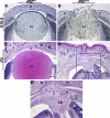
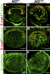
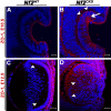
PURPOSE. Neurofibromatosis type 2 (NF2) is an autosomal-dominant CNS tumor syndrome that affects 1:25,000 children and young adults. More than 50% of NF2 patients also develop posterior subcapsular cataracts (PSCs). The authors deleted Nf2 from the lens to determine its role in fiber cell differentiation. METHODS. Nf2 was conditionally deleted from murine lenses using the LeCre transgene. Standard histology and immunohistochemical and immunofluorescent methods were used to analyze lens morphology and markers of cell cycle progression, differentiation, and cell junctions in wild-type and knockout lenses from embryonic day 10.5 through postnatal day 3. RESULTS. Fiber cells lacking Nf2 did not fully exit the cell cycle and continued to express epithelial cell markers, such as FoxE3 and E-cadherin, despite expressing the fiber cell marker Prox1. Many fiber cells lost their elongated morphology. Markers of apical-basal polarity, such as ZO-1, were mislocalized along the lateral and basal membranes of fiber cells. The lens vesicle failed to separate from the surface ectoderm, and prospective lens and corneal epithelial cells formed a multilayered mass of cells at the surface of the eye. Herniation of this membrane caused the fiber mass to erupt through the cornea. CONCLUSIONS. Nf2 is required for complete fiber cell terminal differentiation, maintenance of cell polarity, and separation of lens vesicle from corneal epithelium. Defects identified in fiber cell differentiation may explain the formation of PSCs in patients with NF2. The lens provides an assay system to identify pathways critical for fiber cell differentiation and to test therapies for the tumors that occur in patients with NF2.
Figures


Similar articles
-
Atypical Cadherin Fat1 Is Required for Lens Epithelial Cell Polarity and Proliferation but Not for Fiber Differentiation.Invest Ophthalmol Vis Sci. 2015 Jun;56(6):4099-107. doi: 10.1167/iovs.15-17008. Invest Ophthalmol Vis Sci. 2015. PMID: 26114487 Free PMC article.
-
Spry1 and Spry2 are necessary for lens vesicle separation and corneal differentiation.Invest Ophthalmol Vis Sci. 2011 Aug 29;52(9):6887-97. doi: 10.1167/iovs.11-7531. Invest Ophthalmol Vis Sci. 2011. PMID: 21743007 Free PMC article.
-
Differential requirement for beta-catenin in epithelial and fiber cells during lens development.Dev Biol. 2008 Sep 15;321(2):420-33. doi: 10.1016/j.ydbio.2008.07.002. Epub 2008 Jul 9. Dev Biol. 2008. PMID: 18652817
-
Cataract mutations and lens development.Prog Retin Eye Res. 1999 Mar;18(2):235-67. doi: 10.1016/s1350-9462(98)00018-4. Prog Retin Eye Res. 1999. PMID: 9932285 Review.
-
A role for Wnt/planar cell polarity signaling during lens fiber cell differentiation?Semin Cell Dev Biol. 2006 Dec;17(6):712-25. doi: 10.1016/j.semcdb.2006.11.005. Epub 2006 Nov 19. Semin Cell Dev Biol. 2006. PMID: 17210263 Free PMC article. Review.
Cited by
-
Intrinsic and extrinsic regulatory mechanisms are required to form and maintain a lens of the correct size and shape.Exp Eye Res. 2017 Mar;156:34-40. doi: 10.1016/j.exer.2016.04.009. Epub 2016 Apr 21. Exp Eye Res. 2017. PMID: 27109030 Free PMC article. Review.
-
Defect of LSS Disrupts Lens Development in Cataractogenesis.Front Cell Dev Biol. 2021 Dec 2;9:788422. doi: 10.3389/fcell.2021.788422. eCollection 2021. Front Cell Dev Biol. 2021. PMID: 34926465 Free PMC article.
-
Case series: Pyramidal cataracts, intact irides and nystagmus from three novel PAX6 mutations.Am J Ophthalmol Case Rep. 2018 Feb 28;10:172-179. doi: 10.1016/j.ajoc.2018.02.021. eCollection 2018 Jun. Am J Ophthalmol Case Rep. 2018. PMID: 29780932 Free PMC article.
-
The tumor suppressor gene Trp53 protects the mouse lens against posterior subcapsular cataracts and the BMP receptor Acvr1 acts as a tumor suppressor in the lens.Dis Model Mech. 2011 Jul;4(4):484-95. doi: 10.1242/dmm.006593. Epub 2011 Apr 18. Dis Model Mech. 2011. PMID: 21504908 Free PMC article.
-
Nf2 fine-tunes proliferation and tissue alignment during closure of the optic fissure in the embryonic mouse eye.Hum Mol Genet. 2020 Dec 18;29(20):3373-3387. doi: 10.1093/hmg/ddaa228. Hum Mol Genet. 2020. PMID: 33075808 Free PMC article.
References
-
- Saito H, Yoshida T, Yamazaki H, Suzuki N. Conditional N-rasG12V expression promotes manifestations of neurofibromatosis in a mouse model. Oncogene 2007;26:4714–4719 - PubMed
-
- Gautreau A, Manent J, Fievet B, Louvard D, Giovannini M, Arpin M. Mutant products of the NF2 tumor suppressor gene are degraded by the ubiquitin-proteasome pathway. J Biol Chem 2002;277:31279–31282 - PubMed
-
- McClatchey AI. Neurofibromatosis. Annu Rev Pathol 2007;2:191–216 - PubMed
Publication types
MeSH terms
Substances
Grants and funding
LinkOut - more resources
Full Text Sources
Molecular Biology Databases
Research Materials
Miscellaneous

