. 2010 Apr 1;184(7):3433-41.
doi: 10.4049/jimmunol.0904028. Epub 2010 Feb 24.

Expression of Helios, an Ikaros transcription factor family member, differentiates thymic-derived from peripherally induced Foxp3+ T regulatory cells

Affiliations

Expression of Helios, an Ikaros transcription factor family member, differentiates thymic-derived from peripherally induced Foxp3+ T regulatory cells

Angela M Thornton et al. J Immunol. .

Abstract

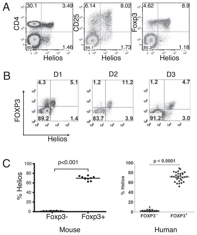
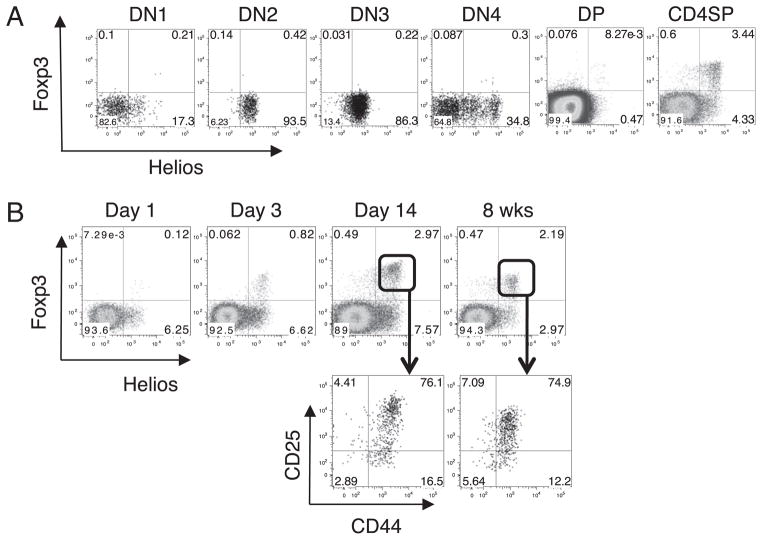
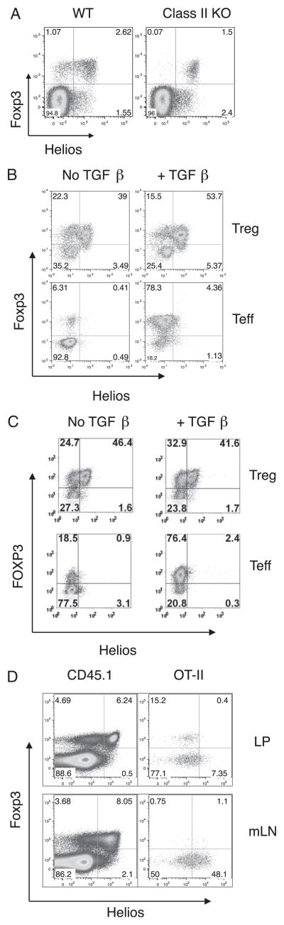
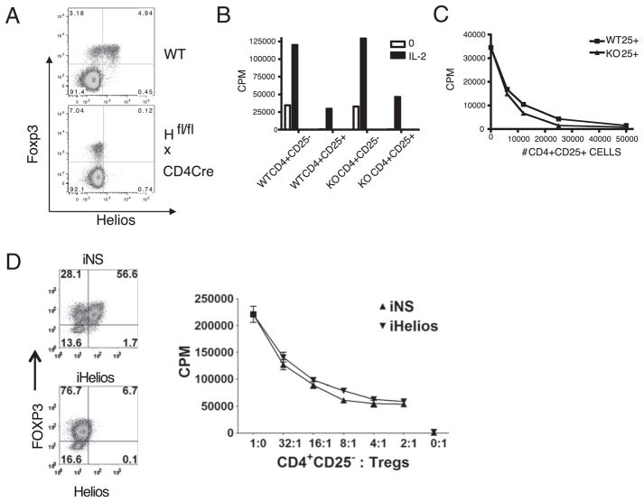
Helios, a member of the Ikaros transcription factor family, is preferentially expressed at the mRNA level by regulatory T cells (Treg cells). We evaluated Helios protein expression using a newly generated mAb and demonstrated that it is expressed in all thymocytes at the double negative 2 stage of thymic development. Although Helios was expressed by 100% of CD4(+)CD8(-)Foxp3(+) thymocytes, its expression in peripheral lymphoid tissues was restricted to a subpopulation ( approximately 70%) of Foxp3(+) T cells in mice and humans. Neither mouse nor human naive T cells induced to express Foxp3 in vitro by TCR stimulation in the presence of TGF-beta expressed Helios. Ag-specific Foxp3(+) T cells induced in vivo by Ag feeding also failed to express Helios. Collectively, these results demonstrate that Helios is potentially a specific marker of thymic-derived Treg cells and raises the possibility that a significant percentage of Foxp3(+) Treg cells are generated extrathymically.


Conflict of interest statement

Disclosures

The authors have no financial conflicts of interest.

Figures

FIGURE 1
FIGURE 1
Helios is expressed in a subset of CD4+Foxp3+ cells. A, Mouse LN cells were analyzed by flow cytometry for Helios expression using mAb 22F6 (left panel). LN cells were gated on CD4+ cells and examined for CD25 and Helios expression (middle panel) and Foxp3 and Helios expression (right panel). B, PBMCs from normal human donors were gated on CD4+ T cells and analyzed for FOXP3 and Helios expression. Three representative FACS plots are shown. C, The percentage of Helios expression in CD4+Foxp3 and CD4+Foxp3 T cells in normal mouse LN or in PBMC of healthy adults. Analysis was performed a minimum of three times with samples from at least two independent mice.
FIGURE 2
FIGURE 2
Expression of Helios during thymic T cell development. A, Mouse thymocytes were stained for CD4, CD8, CD44, and CD25. DN, DP, and CD4SP cells were gated based on CD4 and CD8 expression and were further analyzed by flow cytometry for Helios and Foxp3. DN1–DN4 cells were gated on CD4CD8 cells and further gated based on CD44 and CD25 expression. B, Thymocytes from two individual mice at the indicated ages were analyzed by flow cytometry for Helios and Foxp3 expression. Plots shown are gated on CD4+CD8 thymocytes. Mice were analyzed on different days but were always compared with an adult mouse. Shown are representative plots of one mouse from at least two independent experiments.
FIGURE 3
FIGURE 3
Expression of Helios during peripheral T cell development. A, Splenocytes from two individual mice at the indicated ages were analyzed by flow cytometry for Helios and Foxp3 expression. Plots shown are gated on CD4+ cells. B, Splenocytes from two individual adult or 4-d-old mice were analyzed by flow cytometry for CD25 and CD44 expression. Plots shown are gated on CD4+ cells. C, Splenocytes from two individual mice at the indicated ages were analyzed by flow cytometry for CD25 and CD44 expression. Plots shown are gated on CD4+ Foxp3+Helios cells. Mice of different ages were analyzed on different days but were always compared with an adult mouse. Shown are representative plots of one mouse from at least two independent experiments.
FIGURE 4
FIGURE 4
Thymic-derived Treg cells maintain Helios and Foxp3 expression. CD4+CD8GFP+ thymocytes were sorted from Foxp3-GFP mice, and 6 × 10⁵ cells were adoptively transferred into congenic B6.SJL mice. Four weeks after transfer, LN and spleen cells were analyzed by flow cytometry. Shown are representative profiles of two independent experiments after gating on CD4 and CD45.1+ donor-derived cells.
FIGURE 5
FIGURE 5
Peripherally induced Treg cells are Foxp3+Helios. A, LN cells from two adult C57BL/6 or two MHC class II-deficient mice were analyzed flow cytometry. Plots shown are gated on CD4+ cells and analyzed by flow cytometry for Helios and Foxp3 expression. Shown are representative plots of one mouse. B, CD4+GFP+ or CD4+GFP cells were purified from the LN of Foxp3-GFP mice by cell sorting. Cells were stimulated with plate-bound anti-CD3 and IL-2 in the presence or absence of TGF-β and then analyzed by flow cytometry for Foxp3 and Helios expression. Plots shown are gated on CD4+ cells. Shown is a representative experiment from three independent experiments. C, Human PBMCs were cell sorted for CD4+CD25hiCD127 (Treg) cells and CD4+CD25CD127+ CD45RA+ (Teff) cells. The cells were stimulated in the presence or absence of TGF-β and then analyzed by flow cytometry for Foxp3 and Helios expression. Plots shown are gated on CD4+ cells. Shown is a representative experiment from at least three independent experiments. D, CD4+GFP cells were sorted from OT-II × Foxp3-GFP mice, and 2 × 10⁶ cells were transferred to congenic B6.SJL recipient mice. The recipient mice were given OVA-supplemented drinking water for 6 d. LP cells were isolated and analyzed for Foxp3 and Helios expression by flow cytometry. Plots shown are gated on CD4+CD45.1+ host cells or CD4+CD45.2+ cells (OT-II). A representative result of four individual mice is shown from two independent experiments.
FIGURE 6
FIGURE 6
FOXP3+Helios T cells are enriched in cytokine-producing cells. PBLs from healthy adults were activated for 5 h with PMA/ionomycin, gated on CD4+Foxp3+ cells, and analyzed for Helios and cytokine expression. Panels on the left show a representative plot of Helios versus the indicated cytokine, whereas panels on the right show cumulative data from five healthy adults.
FIGURE 7
FIGURE 7
Helios-deficient Treg cells express Foxp3 and are anergic and suppressive. A, LN cells from two adult C57BL/6 or two Ikzf2fl/fl × CD4-Cre mice were analyzed by flow cytometry. Cells were gated on CD4+ cells and analyzed for Helios and Foxp3 expression. Shown are representative plots of one mouse from three independent experiments. B, CD4+CD25 and CD4+CD25+ cells were isolated from LN of WT C57BL/6 or Ikzf2fl/fl × CD4-Cre mice. Cells were stimulated with T-depleted splenocytes and anti-CD3 in the presence or absence of IL-2 for 3 d. C, WT CD4+CD25 cells were cocultured with the indicated number and type of CD4+CD25+ cells and were stimulated with T-depleted splenocytes and anti-CD3 for 3 d. D, Human Treg cells were activated and expanded for 14 d and then treated with nonspecific (iNS) or Helios (iHelios) siRNA and rested for 24 h. Treated Treg cells were then cultured alone or in titrated numbers with CD4+CD25responders stimulated with HLA-DR+ APCs and soluble anti-CD3 for 3 d before pulsing with [3H]thymidine deoxyribose. Panels on the left represent analysis of Foxp3 and Helios in Treg cells at the end of the coculture. The experiments were set up in triplicate, and a representative experiment from three independent experiments is shown (BD).

Comment in

Similar articles

Cited by

References

    1. Shevach EM. Mechanisms of foxp3+ T regulatory cell-mediated suppression. Immunity. 2009;30:636–645. - PubMed
    1. Josefowicz SZ, Rudensky A. Control of regulatory T cell lineage commitment and maintenance. Immunity. 2009;30:616–625. - PMC - PubMed
    1. Curotto de Lafaille MA, Lafaille JJ. Natural and adaptive foxp3+ regulatory T cells: more of the same or a division of labor? Immunity. 2009;30:626–635. - PubMed
    1. Bennett CL, Christie J, Ramsdell F, Brunkow ME, Ferguson PJ, Whitesell L, Kelly TE, Saulsbury FT, Chance PF, Ochs HD. The immune dysregulation, polyendocrinopathy, enteropathy, X-linked syndrome (IPEX) is caused by mutations of FOXP3. Nat Genet. 2001;27:20–21. - PubMed
    1. Wildin RS, Ramsdell F, Peake J, Faravelli F, Casanova JL, Buist N, Levy-Lahad E, Mazzella M, Goulet O, Perroni L, et al. X-linked neonatal diabetes mellitus, enteropathy and endocrinopathy syndrome is the human equivalent of mouse scurfy. Nat Genet. 2001;27:18–20. - PubMed

Publication types

MeSH terms