. 2010 Apr 1;184(7):3955-63.
doi: 10.4049/jimmunol.0903008. Epub 2010 Feb 24.

Matrix metalloproteinase 12-deficiency augments extracellular matrix degrading metalloproteinases and attenuates IL-13-dependent fibrosis

Affiliations

Matrix metalloproteinase 12-deficiency augments extracellular matrix degrading metalloproteinases and attenuates IL-13-dependent fibrosis

Satish K Madala et al. J Immunol. .

Abstract

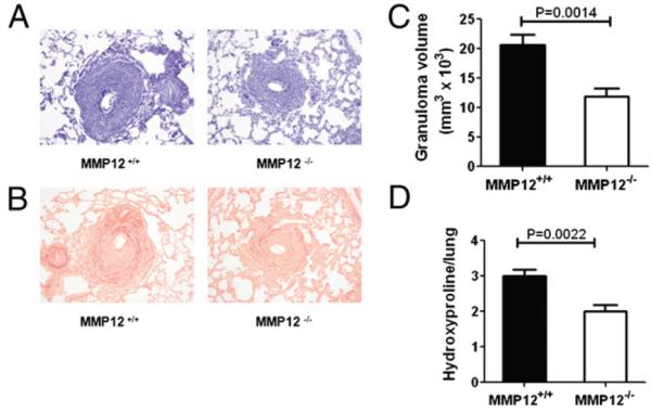
Infection with the parasitic helminth Schistosoma mansoni causes significant liver fibrosis and extracellular matrix (ECM) remodeling. Matrix metalloproteinases (MMP) are important regulators of the ECM by regulating cellular inflammation, extracellular matrix deposition, and tissue reorganization. MMP12 is a macrophage-secreted elastase that is highly induced in the liver and lung in response to S. mansoni eggs, confirmed by both DNA microarray and real-time PCR analysis. However, the function of MMP12 in chronic helminth-induced inflammation and fibrosis is unclear. In this study, we reveal that MMP12 acts as a potent inducer of inflammation and fibrosis after infection with the helminth parasite S. mansoni. Surprisingly, the reduction in liver and lung fibrosis in MMP12-deficient mice was not associated with significant changes in cytokine, chemokine, TGF-beta1, or tissue inhibitors of matrix metalloproteinase expression. Instead, we observed marked increases in MMP2 and MMP13 expression, suggesting that Mmp12 was promoting fibrosis by limiting the expression of specific ECM-degrading MMPs. Interestingly, like MMP12, MMP13 expression was highly dependent on IL-13 and type II-IL-4 receptor signaling. However, in contrast to MMP12, expression of MMP13 was significantly suppressed by the endogenous IL-13 decoy receptor, IL-13Ralpha2. In the absence of MMP12, expression of IL-13Ralpha2 was significantly reduced, providing a possible explanation for the increased IL-13-driven MMP13 activity and reduced fibrosis. As such, these data suggest important counter-regulatory roles between MMP12 and ECM-degrading enzymes like MMP2, MMP9, and MMP13 in Th2 cytokine-driven fibrosis.


Figures

FIGURE 1
FIGURE 1
MMP12 augments S. mansoni egg induced inflammation and fibrosis in liver. C57BL/6 (WT) and mmp12−/− mice were infected with ~30 infective cercariae of S. mansoni and euthanized after 9 wk. Liver tissues were fixed in Bouin-Hollande solution and stained with Giemsa or picrosirius red stain. Average granuloma volumes (A) and liver hydroxyproline levels normalized to egg count (B) are shown. Representative granulomas from WT and mmp12−/− stained with giemsa (C) and picrosirius red (D) are shown at 20× magnification. Statistical significance for data in (A) and (B) is measured using unpaired Student t test (n = 14 per group). The above data are a cumulative representation of two independent experiments that produced similar results.
FIGURE 2
FIGURE 2
MMP12 has no effect on TIMP and cytokines but negatively regulates collagenolytic MMPs. Liver leukocytes isolated from 9 wk S. mansoni-infected mice were separated, counted, and cultured with PMA, Ionomycin, and brefeldin A for 3 h and analyzed for ex vivo cytokine production capability. A, IFN-γ, IL-4, IL-5, and IL-13–producing % cytokine and positive lymphocytes. B, Magnitude of cytokine production by lymphocytes displayed as geometric mean of fluorescence intensity. No statistical significance was observed between the groups (n = 4 per group). RT-PCR analysis was performed for (C) timp1 and (D) timp2 expression in liver tissues at 9 wk infection. Values were normalized to hprt, and fold changes in WT and mmp12−/− were generated by comparing with WT and mmp12−/− unchallenged mice, respectively. The data show the means ± SEM. Statistical significance for data in (C) and (D) is measured using one-way ANOVA (n = 8 per group). E, Liver tissues at wk 9 postinfection were homogenized and soluble lysates (25 μg per well) were subjected to gelatin zymography. Purified mouse MMP2, MMP9, and protein standards were used to identify inactive and active MMP on zymograms and are shown with the arrows in the panel above. F, Immunoblot analysis of liver lysates at 9 wk of S. mansoni infection shows induction of active MMP13 (48 kDa and 20 kDa) that is significantly elevated in MMP12-deficient mice. G, Collagenolytic activity in tissues was measured using FRET peptides that mimic MMP13 target sequences and an increase in the relative fluorescence units (RFUs) depicts MMP activity in samples. Tissue lysates from WT and MMP12−/− were incubated with the reaction mixture containing MMP13 substrate for 30 min and fluorescence was measured using a 96-well fluorescence readers. Statistical significance for data are measured using unpaired Student t test (n = 5 per group). All experiments were repeated twice with similar results.
FIGURE 3
FIGURE 3
MMP12 augments S. mansoni egg induced pulmonary inflammation and fibrosis. S. mansoni egg challenged lungs from WT and mmp12−/− mice were fixed in Bouin-Hollande solution and stained with Giemsa or picrosirius red stain. A, Representative granulomas from WT and mmp12−/− stained with Giemsa (A) or picrosirius red (B) are shown at 20× magnification. C, Reduced granuloma volume in mmp12-deficient mice. D, Reduced lung hydroxyproline levels in mmp12−/− mice on d 8 after eggs. The data show the means ± SEM; significant differences are noted in the figure. Representative of three independent experiments (n = 5–7 per group).
FIGURE 4
FIGURE 4
MMP12 negatively regulates MMP13 expression and pulmonary fibrosis. WT and mmp12−/− mice were sensitized and challenged with S. mansoni eggs. A, After 8 d, RT-PCR analysis was performed in the lungs of WT and mmp12−/− mice for genes mmp12, il-4, il-13, timp1, timp2, and mmp13. Statistical significance for data were measured using one-way ANOVA (n = 6–10 per group). B, Collagenolytic activity in the BAL fluid was measured using FRET peptides that mimic MMP13 target sequences and increase in the relative fluorescence units (RFUs) because of MMP activity in samples. All experiments were repeated twice with similar results. Statistical significance was measured using unpaired Student t test (n = 5 per group).
FIGURE 5
FIGURE 5
MMP13 expression is dependent on IL-13 and its receptors. A, WT, il-10/il-4−/− and il-10/il-12−/− mice were challenged with S. mansoni eggs and euthanized on d 8 and d 15. Transcripts for mmp12 and mmp13 were measured by normalizing to naive mice and the data show the means ± SEM (n = 4 per group). B, WT and IL-13Rα1−/− mice were challenged with S. mansoni eggs and euthanized on d 4 and d 8. Transcripts for mmp12 and mmp13 were measured by normalizing to naive mice and the data show the means ± SEM (n = 5 per group). C, WT and IL-13Rα2−/− mice were challenged with S. mansoni eggs and euthanized on d 4, d 8, and d 15. Transcripts for mmp12 and mmp13 were measured by normalizing to naive mice and the data show the means ± SEM (n = 4 per group). D, After 8 d post eggs, RT-PCR analysis was performed in the lungs of WT and mmp12−/− mice for IL-13Rα2. Significant differences were noted between the groups and the data show the means ± SEM (n = 5 per group). E, RT-PCR analysis in the livers of WT and mmp12−/− mice infected for 9 wk with S. mansoni show reduced IL-13Rα2 in mmp12−/−. The data show the means ± SEM (n = 5 per group). All experiments were repeated twice with similar results.
FIGURE 6
FIGURE 6
MMP13 producing cells in the livers of S. mansoni infected mice. WT and mmp12−/− mice infected with S. mansoni for 9 wk were euthanized, livers were snap frozen and embedded in OCT. A, Liver cryosections were permeabilized and stained for MMP13 (red) and macrophage specific F4/80 (green). Overlaid image shows nuclei stained with DAPI (blue) and rightmost panel shows MMP13 colocalized with F4/80 (white). Scale bar, 100 μm. B, Liver cryosections were permeabilized and stained for MMP13 (green) and fibroblast specific marker α-SMA (red). Overlaid image show nuclei stained with DAPI (blue) and rightmost panel shows MMP13 colocalized with SMA (white). Scale bar, 100 μm. The panels shown are representative of two independent experiments.
FIGURE 7
FIGURE 7
Model of MMP12 in the regulation of S. mansoni-induced fibrosis. Diagram showing proposed action of IL-13–induced MMP12 and matrix degrading MMPs in S. mansoni egg-induced fibrosis.

References

    1. Wynn TA. Common and unique mechanisms regulate fibrosis in various fibroproliferative diseases. J. Clin. Invest. 2007;117:524–529. - PMC - PubMed
    1. Han YP. Matrix metalloproteinases, the pros and cons, in liver fibrosis. J. Gastroenterol. Hepatol. 2006;21(Suppl 3):S88–S91. - PMC - PubMed
    1. Tallant C, Marrero A, Gomis-Ruth FX. Matrix metalloproteinases: Fold and function of their catalytic domains. Biochim Biophys Acta - Molecular. Cell Res. 2010;1803:20–28. - PubMed
    1. Vaillant B, Chiaramonte MG, Cheever AW, Soloway PD, Wynn TA. Regulation of hepatic fibrosis and extracellular matrix genes by the th response: new insight into the role of tissue inhibitors of matrix metalloproteinases. J. Immunol. 2001;167:7017–7026. - PubMed
    1. Wilson MS, Mentink-Kane MM, Pesce JT, Ramalingam TR, Thompson R, Wynn TA. Immunopathology of schistosomiasis. Immunol. Cell Biol. 2007;85:148–154. - PMC - PubMed

Publication types

MeSH terms