. 2010 May;298(5):C1198-208.
doi: 10.1152/ajpcell.00511.2009. Epub 2010 Feb 24.

Extracellular acidosis activates ASIC-like channels in freshly isolated cerebral artery smooth muscle cells

Affiliations

Extracellular acidosis activates ASIC-like channels in freshly isolated cerebral artery smooth muscle cells

Wen-Shuo Chung et al. Am J Physiol Cell Physiol. 2010 May.

Abstract

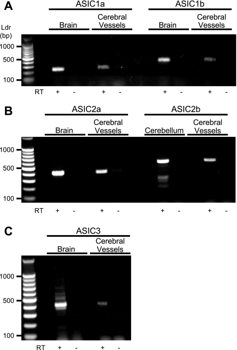
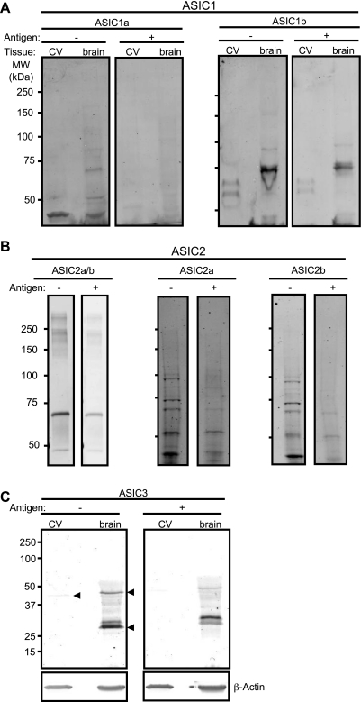
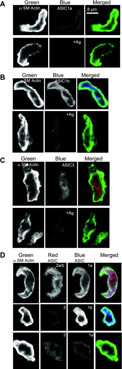
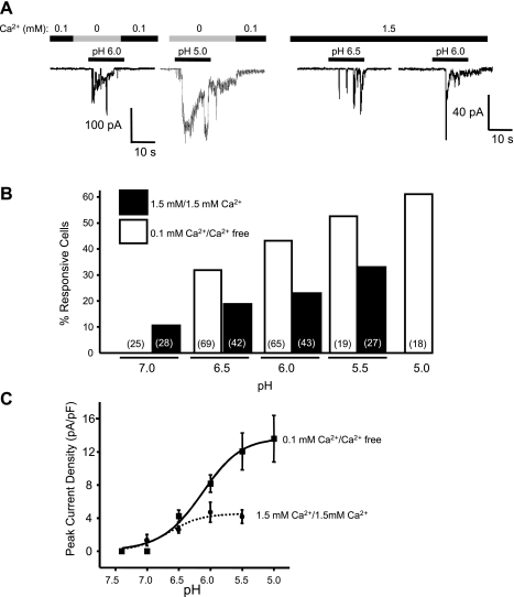
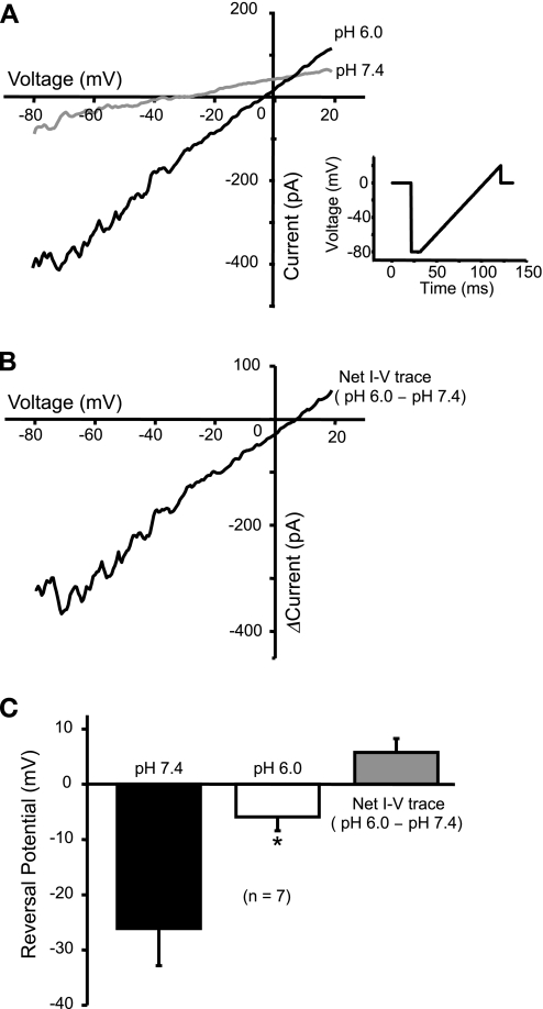
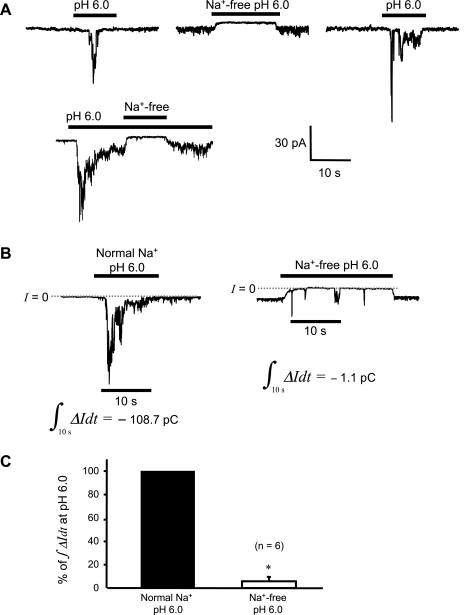
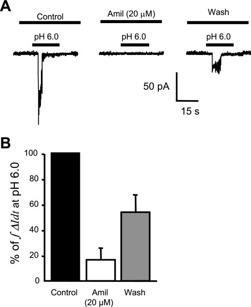
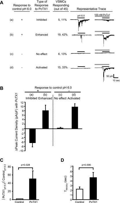
Recent studies suggest that certain acid-sensing ion channels (ASIC) are expressed in vascular smooth muscle cells (VSMCs) and are required for VSMC functions. However, electrophysiological evidence of ASIC channels in VSMCs is lacking. The purpose of this study was to test the hypothesis that isolated cerebral artery VSMCs express ASIC-like channels. To address this hypothesis, we used RT-PCR, Western blotting, immunolabeling, and conventional whole cell patch-clamp technique. We found extracellular H(+)-induced inward currents in 46% of cells tested (n = 58 of 126 VSMCs, pH 6.5-5.0). The percentage of responsive cells and the current amplitude increased as the external H(+) concentration increased (pH(6.0), n = 28/65 VSMCs responsive, mean current density = 8.1 +/- 1.2 pA/pF). Extracellular acidosis (pH(6.0)) shifted the whole cell reversal potential toward the Nernst potential of Na(+) (n = 6) and substitution of extracellular Na(+) by N-methyl-d-glucamine abolished the inward current (n = 6), indicating that Na(+) is a major charge carrier. The broad-spectrum ASIC blocker amiloride (20 microM) inhibited proton-induced currents to 16.5 +/- 8.7% of control (n = 6, pH(6.0)). Psalmotoxin 1 (PcTx1), an ASIC1a inhibitor and ASIC1b activator, had mixed effects: PcTx1 either 1) abolished H(+)-induced currents (11% of VSMCs, 5/45), 2) enhanced or promoted activation of H(+)-induced currents (76%, 34/45), or 3) failed to promote H(+) activation in nonresponsive VSMCs (13%, 6/45). These findings suggest that freshly dissociated cerebral artery VSMCs express ASIC-like channels, which are predominantly formed by ASIC1b.


Figures

Fig. 1.
Fig. 1.
RT-PCR detection of acid-sensing ion channel (ASIC) mRNA expression in mouse cerebral vessels. A: ASIC1. B: ASIC2. C: ASIC3. Whole brain or cerebellum was used as a positive control where noted. The presence of reverse transcriptase is indicated (RT+). Samples without RT served as negative controls. Base pair ladder (Ldr) is shown at left.
Fig. 2.
Fig. 2.
Western blot detection of ASIC proteins in isolated cerebral vessels (CV). Representative blots for ASIC1a and ASIC1b (A), ASIC2a/b, ASIC2a, and ASIC2b (B), and ASIC3 (C) are shown. β-Actin was used as a loading control for ASIC3. +/− denotes presence/absence of antigen. Arrowheads in C identify bands that are diminished in corresponding antigen control. MW, molecular weight.
Fig. 3.
Fig. 3.
Immunolocalization of ASIC proteins in isolated cerebral vascular smooth muscle cells (VSMCs). Dissociated cerebral VSMCs were labeled for α-smooth muscle (α-SM) actin (far left in white, green in merged image on right), a VSMC marker, and ASIC proteins. AC: representative examples of antibody and antibody + antigen (+Ag) controls for ASIC1a (A), ASIC1b (B), and ASIC3 (C). D: VSMCs labeled for ASIC2a/b and ASIC1a, ASIC3 and ASIC1b, and ASIC3 and ASIC1a. Nearly all VSMCs examined contained labeling for ASIC1a (14/16), ASIC1b (13/13), ASIC2a/b (31/31), and ASIC3 (13/14).
Fig. 4.
Fig. 4.
A: shape of a representative isolated cerebral VSMC. B: protocol for exposure to extracellular protons. VSMCs were briefly exposed to nominally Ca2+-free solution before exposure to extracellular H+ to prevent contraction. C and D: representative traces of the two main types of extracellular H+-evoked currents found in cerebral VSMCs.
Fig. 5.
Fig. 5.
pH dependence of acid-induced inward current in freshly isolated cerebral VSMCs. A: representative traces of currents evoked by various concentrations of H+ ([H+]) when conditioned with 0.1 mM (left) and 1.5 mM Ca2+ (right). B: a tendency of pH dependence was also seen in the percentage of cells exhibiting acid-induced inward currents. C: plot of pH-current relation shows pH dependence of the peak current density (peak currents normalized to their respective membrane capacitances) in responsive cells. A pH-response curve was obtained by fitting the data to the Hill equation. The pH50 was 6.15 and 6.65 for conditioning with 0.1 and 1.5 mM Ca2+, respectively.
Fig. 6.
Fig. 6.
Current-voltage (I-V) relation changed by extracellular acidosis. A: I-V curves in pH 7.4 and pH 6.0 were acquired using a voltage-ramp protocol. Exposure to pH 6.0 increased the slope of I-V curve, indicating an increase in conductance during acidosis. The whole cell reversal potential (when I = 0) is also shifted to the right toward equilibrium potential of Na+ (ENa). Inset A: voltage-ramp protocol and waveform used in this experiment to acquire I-V curve. B: the net effect of pH 6.0 on I-V curve is shown, which is obtained by deducting the curve at pH 7.4 from that at pH 6.0. C: statistic shows a significant shift of the whole cell reversal potential to the right (*P < 0.05). The average net reversal potential is slightly positive (n = 5).
Fig. 7.
Fig. 7.
H+-gated inward current is dependent on external Na+. A: segments from a representative continuous recording, showing that removal of external Na+ obliterated current induced by pH 6.0. B: integration of the inward current over a 10-s time interval during acid exposure gives the amount of charge transported during that interval. The interval picked starts from 2 s before the peak current. A substantial reduction in charge movement is seen in the example shown (pC = pA·s). C: the average charge movement in the absence of external Na+ is significantly reduced to only 5.8 ± 3.9% of the control, a decrease of >94% (*P < 0.05).
Fig. 8.
Fig. 8.
Pharmacological inhibition of acid-induced currents by the broad-spectrum ASIC inhibitor amiloride (Amil) in isolated cerebral VSMCs. Representative trace (A) and group data in response to 20 μM amiloride (B) are shown. Cells were pretreated with the amiloride 1 min before and during the second exposure to pH 6.0.
Fig. 9.
Fig. 9.
Effect of psalmotoxin 1 (PcTx1), an ASIC1a inhibitor and ASIC1b activator, on H+-induced currents in isolated cerebral VSMCs. A: representative traces of acid-induced current response to preconditioning with 100 nM PcTx1 for 30 s. Traces of VSMCs (24 out of 45) that responded to the control H+ stimulus are shown in a and b. In these cells, inward currents were either inhibited (a) or enhanced (b). Currents were enhanced (19/45) nearly four times as often as they were inhibited (5/45). Traces of VSMCs (21/45) that did not respond to an initial extracellular H+ stimulus are shown in c and d. In these cells, PcTx1 had no effect (c) or resulted in activation of currents (d). Currents were activated (15/45) nearly twice as often as they were unaffected (6/45). Thus, PcTx1 predominantly potentiated H+-gated currents in VSMCs. B: group data summarizing the effect of PcTx1 on the change in peak current density. C and D: in the VSMCs where PcTx1 enhanced or activated current, PcTx1 increased total current density relative to control (∫PcTx1pH6.0/∫controlpH6.0) (C) and delayed desensitization (τdecay, decay time constant) of the inward current (D).

References

    1. Babini E, Paukert M, Geisler HS, Grunder S. Alternative splicing and interaction with di- and polyvalent cations control the dynamic range of acid-sensing ion channel 1 (ASIC1). J Biol Chem 277: 41597–41603, 2002 - PubMed
    1. Baron A, Deval E, Salinas M, Lingueglia E, Voilley N, Lazdunski M. Protein kinase C stimulates the acid-sensing ion channel ASIC2a via the PDZ domain-containing protein PICK1. J Biol Chem 277: 50463–50468, 2002 - PubMed
    1. Benos DJ, Stanton BA. Functional domains within the degenerin/epithelial sodium channel (Deg/ENaC) superfamily of ion channels. J Physiol 520 Pt 3: 631–644, 1999 - PMC - PubMed
    1. Benson CJ, Xie J, Wemmie JA, Price MP, Henss JM, Welsh MJ, Snyder PM. Heteromultimers of DEG/ENaC subunits form H+-gated channels in mouse sensory neurons. Proc Natl Acad Sci USA 99: 2338–2343, 2002 - PMC - PubMed
    1. Berdiev BK, Xia J, Jovov B, Markert JM, Mapstone TB, Gillespie GY, Fuller CM, Bubien JK, Benos DJ. Protein kinase C isoform antagonism controls BNaC2 (ASIC1) function. J Biol Chem 277: 45734–45740, 2002 - PubMed

Publication types

LinkOut - more resources