. 2010 May;298(5):C1140-50.
doi: 10.1152/ajpcell.00242.2009. Epub 2010 Feb 24.

p38 MAP kinase is necessary for melanoma-mediated regulation of VE-cadherin disassembly

Affiliations

p38 MAP kinase is necessary for melanoma-mediated regulation of VE-cadherin disassembly

Payal Khanna et al. Am J Physiol Cell Physiol. 2010 May.

Abstract

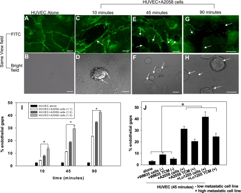
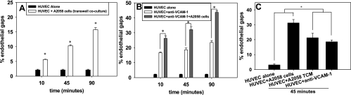
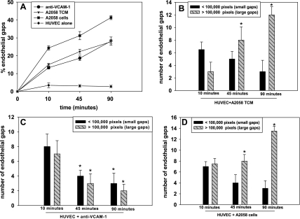
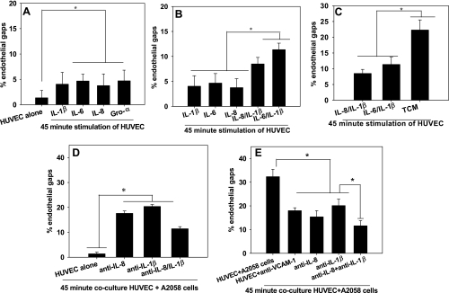
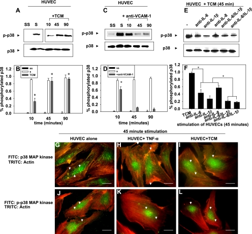
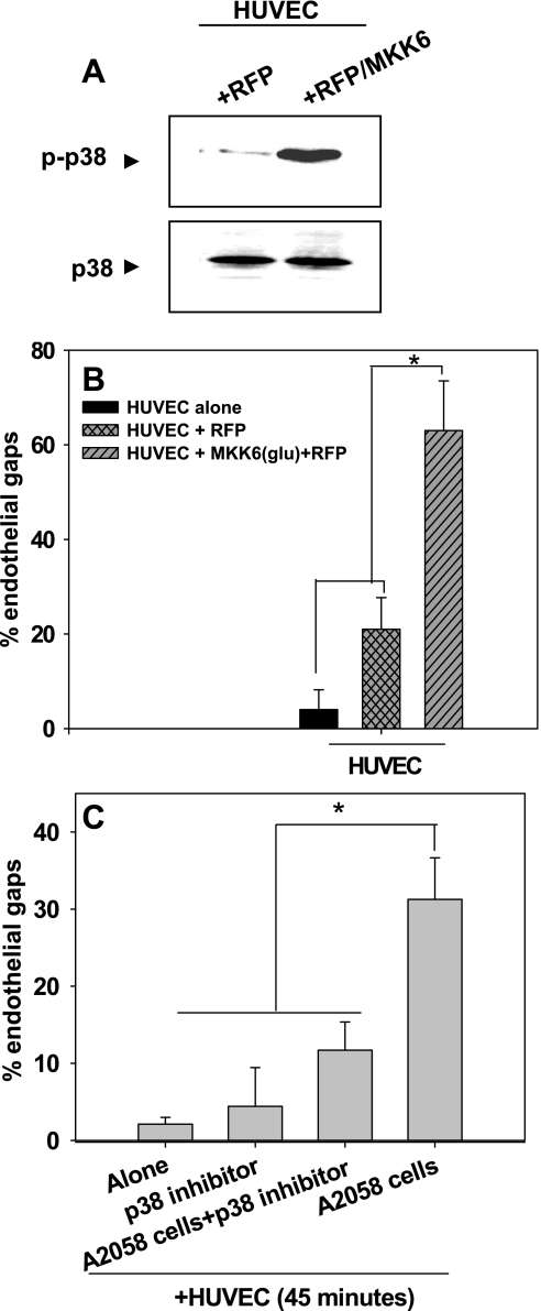
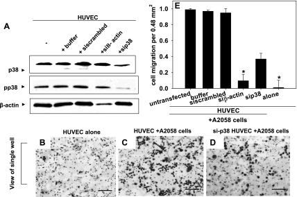
Vascular endothelial (VE)-cadherin is localized to the endothelial borders and the adherens junctions, which are regulated by changes in mitogen-activated protein (MAP) kinases, GTPases, and intracellular calcium. We previously showed that melanoma cells induce VE-cadherin disassembly through contact with human umbilical vein endothelial cells in coculture. However, the exact mechanism by which melanoma cells signal endothelial cells to induce VE-cadherin junction disassembly is not well understood. In this study, VE-cadherin junction disassembly was further examined under fluorescence microscopy. We found that melanoma-induced VE-cadherin junction disassembly and upregulation of p38 MAP kinase in endothelial cells is regulated by both soluble factors from melanomas, particularly interleukin (IL)-8, IL-6, and IL-1beta, and through vascular cell adhesion molecule-1. Neutralizing melanoma-secreted soluble factors reduced endothelial gap formation. Endothelial cells transfected with MAP kinase kinase 6, a direct activator of p38 MAP kinase, increased VE-cadherin-mediated gap formation, facilitating melanoma transendothelial migration. In contrast, endothelial cells transfected with small-interfering RNA against p38 MAP kinase expression largely prevented melanoma transendothelial migration in Boyden chamber experiments. These findings indicate that p38 MAP kinase proteins regulate VE-cadherin junction disassembly, facilitating melanoma migration across endothelial cells.


Figures

Fig. 1.
Fig. 1.
Melanoma cells induce vascular endothelial (VE)-cadherin disassembly. Bars in A–H, 5 μm. A: VE-cadherin junctions in human umbilical vein endothelial cells (HUVECs, 0.3 × 10⁶ cells) without A2058 melanoma cells. HUVECs were fixed, permeabilized, and stained with anti-VE-cadherin monoclonal antibody (mAb) followed by Alexa 488. Representative fields were examined and show intact VE-cadherin junctions indicated by intact green fluorescent borders. B: in the same field of view as A, there are no tumor cells observed. C–H: disruption of VE-cadherin junctions after HUVECs were in direct contact with A2058 tumor cells for either 10, 45, or 90 min are shown using FITC. The same field of view captured under brightfield shows tumor cells in regions coinciding with gap formation. Arrows show disruption of VE-cadherin homodimers. I: HUVECs were cocultured with different concentrations of A2058 melanoma cells for 10, 45, and 90 min at 1:1, 1:2, or 1:3 ratios of HUVECs to A2058 cells. P values compare each experimental condition with %gap of HUVEC alone (*P < 0.05). J: HUVECs were cocultured in direct contact with tumor conditioned medium (TCM) or melanoma cells of increasing metastatic potential for 45 min. P values compare each experimental condition with %gap of HUVEC alone and WM35 cells/TCM (*P < 0.05). For all experiments, values are means ± SD.
Fig. 2.
Fig. 2.
Endothelial gaps increase with metastatic potential of melanoma cells, which are mediated by both soluble factors and anti-vascular cell adhesion molecule-1 (VCAM-1). A: HUVECs were placed in indirect coculture with A2058 melanoma cells for 10, 45, and 90 min. The Transwell insert with melanoma cells was removed and %area of gap was assessed. P values compare each experimental condition with %gap of HUVEC alone (*P < 0.05). B: here, HUVECs were stimulated with anti-VCAM-1 for 45 min before adding A2058 melanoma cells. P values are comparing %endothelial gap of HUVEC + anti-VCAM-1 and HUVEC + anti-VCAM-1 + A2058 cells with %endothelial gap of HUVEC alone (*P < 0.05). C: HUVECs cocultured with TCM or anti-VCAM-1 significantly increase %gap formation compared with HUVECs alone, but less gap formation than A2058 cells cocultured with HUVECs. P values are comparing %endothelial gap of HUVEC + TCM and HUVEC + anti-VCAM-1 with %endothelial gap of HUVEC alone and HUVEC + A2058 cells (*P < 0.05). For all experiments, values are means ± SD.
Fig. 3.
Fig. 3.
Endothelial gap sizes increase linearly over time. Experiments show the %endothelial gaps over time when A2058 TCM, anti-VCAM-1, or A2058 cells are cocultured with HUVECs over 10, 45, or 90 min. A: the %endothelial gaps as a function of time are plotted. B: the no. of gaps above or below an area of 100,000 pixels increased as a function of time; %endothelial gaps was plotted from data collected in A. P values are comparing the no. of large gaps in HUVEC + A2058 TCM after 45 and 90 min with no. of large gaps in HUVEC + A2058 TCM after 10 min (*P < 0.05). C: from data in A, the no. of endothelial gaps above or below an area of 100,000 pixels was plotted over time. P values are comparing the no. of large gaps in HUVEC + anti-VCAM-1 after 45 and 90 min with the number of large gaps in HUVEC + anti-VCAM-1 after 10 min (*P < 0.05). D: from data in A, the no. of small and large endothelial gaps was plotted as a function of time when HUVECs were cocultured with A2058 cells. P values are comparing the no. of large gaps in HUVEC + A2058 cells after 45 and 90 min with the no. of large gaps in HUVEC + A2058 cells after 10 min (*P < 0.05). All values are means ± SD.
Fig. 4.
Fig. 4.
A–C: interleukin (IL)-8 and IL-1β have additive rather than synergistic effects on VE-cadherin disassembly. A: the indicated cytokines induced VE-cadherin disassembly. P values are comparing %gap area of cytokines with %gap of HUVEC alone (*P < 0.05). B: stimulation of HUVECs with recombinant forms of IL-8 and IL-1β or IL-6 and IL-1β induced additive effects on endothelial gap formation. Concentrations of cytokines were based on TCM concentrations measured using ELISA (Table 2). P values compare %gap area of combinations of cytokines with %gap of HUVEC + IL-8, HUVEC + IL-6, and HUVEC + IL-1β. C: combinations of cytokines induced significantly less gap formation than TCM alone. P values are comparing %gap area of TCM with %gap of HUVEC + IL-8/IL-1β and HUVEC + IL-6/ IL-1β. D and E: anti-VCAM-1 and neutralization of both IL-8 and IL-1β dramatically reduces the breakdown of VE-cadherin. D: HUVECs were cocultured with A2058 cells + anti-IL-8, A2058 cells + anti-IL-1β, or A2058 cells + anti-IL-8 + anti-IL-1β. P values are comparing each experimental condition with %endothelial gap areas during HUVEC alone (*P < 0.05). E: using the same controls as in D, data were graphed to make comparisons between the effects of neutralizing individual and pairs of cytokines. HUVECs were cocultured with either A2058 cells, anti-VCAM-1, A2058 cells + anti-IL-8, A2058 cells + anti-IL-1β, or A2058 cells + anti-IL-8 + anti-IL-1β. Neutralization of both IL-8 and IL-1β dramatically decreased %endothelial gaps compared with HUVECs stimulated with anti-VCAM-1 or anti-IL-8 and anti-IL-1β alone. P values are comparing each experimental condition with %endothelial gap areas for HUVEC + anti-VCAM-1, HUVEC + A2058 cells + anti-IL-8, and HUVEC + A2058 cells + anti-IL-1β (*P < 0.05). Values for graphs and ELISA are means ± SD.
Fig. 5.
Fig. 5.
A–F: soluble factors and anti-VCAM-1 regulate p38 phosphorylation. A: Western blots show p38 phosphorylation (p) when HUVECs are stimulated with A2058 TCM for 10, 45 min, and 90 min. The location of “S” and “SS” lanes have been moved for ease of interpretation but are from the same Western blot as other lanes. B: Western blots in A were quantified. P values are comparing TCM-stimulated case with the SS case. C: effects of anti-VCAM-1 on p38 phosphorylation in HUVECs over 10, 45, and 90 min. D: Western blots in C were quantified. P values are comparing TCM-stimulated case with the SS case. E: effects of neutralizing cytokines in TCM from A2058 cells for 45 min. Neutralizing antibodies (indicated) were respectively added in TCM before coculture with HUVECs. F: Western blots in E were quantified. P values are comparing TCM-stimulated case with cases where IL-8, IL-1β, IL-6, or a combination of these were neutralized in TCM and then added to HUVECs. Western blots are representative of 3 different experiments. Values are means ± SD. G–L: p38 mitogen-activated protein (MAP) kinase proteins translocate from the cytosol to the nucleus after phosphorylation (bars, 25 μm). HUVECs were fixed and stained with anti-p38 followed by Alexa 488-labeled secondary antibodies. The actin cytoskeleton was stained with phalloidin conjugated to Alexa 546. G: fluorescent images of unstimulated HUVECs. H: fluorescent images of HUVECs stimulated with recombinant tumor necrosis factor (TNF)-α for 45 min. I: HUVECs in contact with A2058 TCM for 45 min. J: fluorescent images of HUVECs fixed and stained with phospho-p38 followed by Alexa 488 secondary antibodies and phalloidin. K: fluorescent images of HUVECs stimulated with recombinant TNF-α for 45 min. L: fluorescent images of HUVECs in contact with A2058 TCM for 45 min. Images are representative of 3 separate experiments.
Fig. 6.
Fig. 6.
Overexpression of p38 increases disassembly of VE-cadherin homodimers to form endothelial gaps. A: transfection efficiency of HUVECs with red fluorescent protein (RFP) or MAP kinase kinase (MKK) 6(glu)/RFP was assessed using Western blots probed for either anti-p38 or anti-phospho-p38. Western blots are representative of three separate experiments. B: HUVECs were transfected with constitutively active MKK6(glu) fused with RFP to label transfected cells. Endothelial gap formation was assessed 36–48 h posttransfection. P values are comparing each experimental condition with %gap of HUVECs alone and HUVECs + RFP (*P < 0.05). C: inhibiting p38 phosphorylation prevents the disassembly of VE-cadherin homodimers. HUVECs were treated with SB-220025 for 30 min before they were cocultured with A2058 melanoma cells for 45 min. Values are means ± SD. P values are comparing each experimental condition with %gap of HUVEC + A2058 cells (*P < 0.05).
Fig. 7.
Fig. 7.
p38 MAP kinase activation facilitates melanoma extravasation. A: small-interfering (si) RNA knockdown of p38 (and thus pp38) was confirmed using Western blots. HUVECs transfected with siRNA against p38 (sip38), β-actin (siβ-actin), scrambled control, or buffer were lysed and subjected to SDS-PAGE to assess knockdown efficiency. B–D represent images of the underside of the polycarbonate filters from experiments using the Boyden chamber. Bars, 40 μm. B: HUVECs alone without A2058 cells. Bars, 40 μm C: HUVECs plus 50 μl of A2058 tumor cells (0.9 X 10⁶ cells/ml) added to each of the 12 wells on the top plate of the Boyden chamber. D: HUVECs transfected with p38 siRNA plus A2058 tumor cells. The images of A2058 transmigration and Western blots are representative of 3 different experiments. E: average of migrated A2058 tumor cells for each experimental and control case. Values are means ± SD. P values are comparing each experimental condition with A2058 extravasation through untransfected HUVEC (*P < 0.05).

Similar articles

Cited by

References

    1. Alcaide P, Newton G, Auerbach S, Sehrawat S, Mayadas TN, Golan DE, Yacono P, Vincent P, Kowalczyk A, Luscinskas FW. p120-Catenin regulates leukocyte transmigration through an effect on VE-cadherin phosphorylation. Blood 112: 2770–2779, 2008 - PMC - PubMed
    1. Arroyo AG, Sánchez-Mateos P, Campanero MR, Martín-Padura I, Dejana E, Sánchez-Madrid F. Regulation of the VLA integrin-ligand interactions through the beta 1 subunit. J Cell Biol 117: 659–670, 1992 - PMC - PubMed
    1. Dejana E, Bazzoni G, Lampugnani MG. Vascular endothelial (VE)-cadherin: only an intercellular glue? Exp Cell Res 252: 13–19, 1999 - PubMed
    1. Dong C, Slattery MJ, Liang S, Peng HH. Melanoma cell extravasation under flow conditions is modulated by leukocytes and endogenously produced interleukin 8. Mol Cell Biomech 2: 145–159, 2005 - PMC - PubMed
    1. Dong C, Slattery MJ, Rank BM, You J. In vitro characterization and micromechanics of tumor cell chemotactic protrusion, locomotion, and extravasation. Ann Biomed Eng 30: 344–355, 2002 - PMC - PubMed

Publication types

MeSH terms