. 2010 Jul;24(7):2334-46.
doi: 10.1096/fj.09-147991. Epub 2010 Feb 24.

Cockayne syndrome group B protein promotes mitochondrial DNA stability by supporting the DNA repair association with the mitochondrial membrane

Affiliations

Cockayne syndrome group B protein promotes mitochondrial DNA stability by supporting the DNA repair association with the mitochondrial membrane

Maria D Aamann et al. FASEB J. 2010 Jul.

Abstract

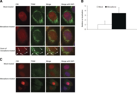
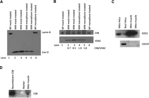
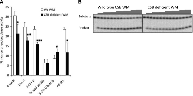
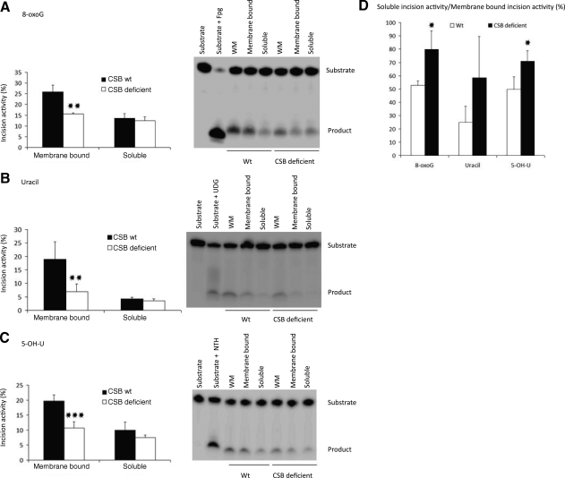
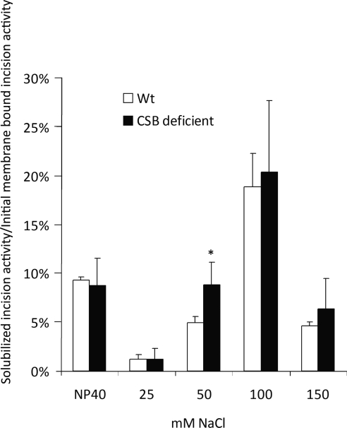
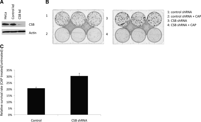
Cockayne syndrome (CS) is a human premature aging disorder associated with severe developmental deficiencies and neurodegeneration, and phenotypically it resembles some mitochondrial DNA (mtDNA) diseases. Most patients belong to complementation group B, and the CS group B (CSB) protein plays a role in genomic maintenance and transcriptome regulation. By immunocytochemistry, mitochondrial fractionation, and Western blotting, we demonstrate that CSB localizes to mitochondria in different types of cells, with increased mitochondrial distribution following menadione-induced oxidative stress. Moreover, our results suggest that CSB plays a significant role in mitochondrial base excision repair (BER) regulation. In particular, we find reduced 8-oxo-guanine, uracil, and 5-hydroxy-uracil BER incision activities in CSB-deficient cells compared to wild-type cells. This deficiency correlates with deficient association of the BER activities with the mitochondrial inner membrane, suggesting that CSB may participate in the anchoring of the DNA repair complex. Increased mutation frequency in mtDNA of CSB-deficient cells demonstrates functional significance of the presence of CSB in the mitochondria. The results in total suggest that CSB plays a direct role in mitochondrial BER by helping recruit, stabilize, and/or retain BER proteins in repair complexes associated with the inner mitochondrial membrane, perhaps providing a novel basis for understanding the complex phenotype of this debilitating disorder.


Figures

Figure 1.
Figure 1.
Colocalization of CSB with TFAM in menadione treated cells. A) HeLa cells either mock treated or treated with 200 μM menadione were stained for endogenous CSB and TFAM. Colocalization in the merged fields appears as yellow foci (arrows). B) Colocalization was quantified using Volocity software. Solid bar, fold increase in colocalization of CSB to TFAM after menadione treatment; open bar, mock treatment. Results from one representative experiment are shown. C) Primary human skin fibroblast cells either mock treated or treated with 200 μM menadione were stained for endogenous CSB and TFAM. Colocalization in merged fields appears as yellow foci.
Figure 2.
Figure 2.
Presence of CSB in whole mitochondrial extract. A, B) Mitochondria were isolated from HeLa cells either mock treated or treated with 200 μM menadione; see Materials and Methods. A) Fifteen micrograms purified whole mitochondria (WM) or nuclear pellet (NP) from both mock and menadione-treated cells were used for Western blot using Lamin B as nuclear marker and mitochondrial protein Cox IV. B) Fifteen micrograms of both NP and WM were used for Western blot, which was cut at 100 kDa; bottom part was probed for structural mitochondrial protein VDAC and top part for CSB. Two separate samples for each treatment (lanes 2, 3 and 4, 5, respectively) of whole mitochondria were loaded on the gel. Relative band intensities of CSB/VDAC, shown below the Western blot, were quantified using ImageQuant TL. C, D) Mitochondria were isolated from primary human muscle cells. C) Top panel: 40-μg nuclear and mitochondrial samples from HeLa cells and 40-μg mitochondrial extract from muscles were used for Western blot against SOD-2. Bottom panel: 60-μg mitochondrial extract from muscles was used for Western blot against CDC47 to test for nuclear contamination with 60-μg nuclear samples from HeLa cells as positive control. D) Purified CSB and muscle mitochondria (50 μg) were used for Western blot against CSB.
Figure 3.
Figure 3.
Purity of mitochondria extract from CS1AN cells. Mitochondria from SV-40 transformed CS1AN cells either nontransfected or stably transfected with either empty vector (vec) or wild-type CSB expression vector (wt) were purified as described in Materials and Methods. Whole mitochondrial extract (WM; 40 μg) or nuclear extract (NE; 25 μg) was resolved on a 12% Tris-glycine gel and transferred to a PVDF membrane. Membrane was probed with antibodies against nuclear protein Lamin B (Novocastra) and mitochondrial protein CoxIV (Santa Cruz), and film was overexposed for the Lamin B signal.
Figure 4.
Figure 4.
Decreased repair activity in CSB-deficient mitochondrial extract. Incision of whole mitochondrial extract (WM) from CS1AN cells stably transfected with either empty vector (CSB-deficient) or CSB wild-type expression vector (wt). A) Incision represented as percentage cleaved substrate containing 8-oxoG, uracil, or 5-OH-U in a double-stranded oligodeoxynucleotide; 8-oxoG or 5-OH-U in an 11-nt bubble; or THF, an AP site analog, in a double-stranded oligodeoxynucleotide (Table 1). Results are means ± sd for 3–4 independent experiments. *P < 0.05, **P < 0.01, ***P < 0.001 vs. wt. B) Representative gel picture for THF incision assay using WM from CSB wild-type cells and CSB-deficient cells, with increasing amounts of mitochondrial extracts from left to right.
Figure 5.
Figure 5.
Design of mitochondrial fractionation procedure. A) Fractionation of whole mitochondria into membrane-bound fraction and soluble fraction, used for experiments shown in Fig. 6. B) Additional subfractionation of membrane-bound fraction 1. Membrane-bound fraction was subjected to Nonidet P-40 treatment, followed by increasing salt concentrations, followed by centrifugation, resulting in soluble fraction 2 to 6, all originating from the initial membrane-bound fraction 1. Soluble fractions were used for experiments shown in Fig. 7.
Figure 6.
Figure 6.
Decreased association of incision capacity for 8-oxoG, uracil, and 5-OH-U with the inner mitochondrial membrane. Mitochondria from CS1AN stably transfected with empty vector (CSB deficient) or wild-type CSB (wt) were isolated and fractionated into a soluble and a membrane-bound fraction. A) Incision activity for 8-oxoG. Right panel: representative gel for incision activity in whole mitochondria (WM), membrane-bound fraction 1, and soluble fraction 1. Left panel: quantified incision activity in the membrane-bound fraction 1 and soluble fraction 1. B) Incision activity for uracil. Right panel: representative gel for incision. Left panel: quantified incision activity. C) Incision activity for 5-OH-U. Right panel: representative gel for incision activity. Left panel: quantified incision activity. D) Soluble fraction 1 incision activity relative to membrane-bound fraction 1 incision activity. Results are means ± sd for 3–4 independent experiments. *P < 0.05, **P < 0.01, ***P < 0.001 vs. wt.
Figure 7.
Figure 7.
Release of uracil incision activity by increasing salt concentration. Mitochondrial proteins were fractionated from membrane-bound fraction 1, as depicted in Fig. 5B. Uracil incision capacity in resulting soluble fractions was measured. Incision activity in soluble fraction relative to incision capacity of membrane-bound fraction 1 is presented. *P < 0.05 vs. wt.
Figure 8.
Figure 8.
Increased mitochondrial mutagenesis in CSB knockdown cells. A) Western blot of 50-μg cell extracts from HeLa cells and HeLa cells either stably transfected with control shRNA or CSB shRNA, as indicated, probed with CSB and actin antibody. CSB knockdown was calculated relative to loading control actin levels. B) Chloramphenicol (CAP) resistance assay. HeLa cells with a stable shRNA CSB knockdown and shRNA control cells were seeded and grown in presence or absence of CAP. C) Numbers of colonies formed with and without 200 μg/ml CAP treatment was quantified, and relative survival rate determined as colonies formed with CAP divided by the numbers of colonies formed without CAP. Results are presented as a representative experiment performed in triplicate.

Similar articles

Cited by

References

    1. Balajee A S, Proietti De Santis L, Brosh R M, Jr, Selzer R, Bohr V A. Role of the ATPase domain of the Cockayne syndrome group B protein in UV induced apoptosis. Oncogene. 2000;19:477–489. - PubMed
    1. Lehmann A R. DNA repair-deficient diseases, xeroderma pigmentosum, Cockayne syndrome and trichothiodystrophy. Biochimie (Paris) 2003;85:1101–1111. - PubMed
    1. Stevnsner T, Nyaga S, de Souza-Pinto N C, van der Horst G T, Gorgels T G, Hogue B A, Thorslund T, Bohr V A. Mitochondrial repair of 8-oxoguanine is deficient in Cockayne syndrome group B. Oncogene. 2002;21:8675–8682. - PubMed
    1. Wong H K, Muftuoglu M, Beck G, Imam S Z, Bohr V A, Wilson D M., 3rd Cockayne syndrome B protein stimulates apurinic endonuclease 1 activity and protects against agents that introduce base excision repair intermediates. Nucleic Acids Res. 2007;35:4103–4113. - PMC - PubMed
    1. Dianov G, Bischoff C, Sunesen M, Bohr V A. Repair of 8-oxoguanine in DNA is deficient in Cockayne syndrome group B cells. Nucleic Acids Res. 1999;27:1365–1368. - PMC - PubMed

Publication types

LinkOut - more resources