. 2010 Apr 23;285(17):13294-303.
doi: 10.1074/jbc.M109.088864. Epub 2010 Feb 24.

Interaction between periostin and BMP-1 promotes proteolytic activation of lysyl oxidase

Affiliations

Interaction between periostin and BMP-1 promotes proteolytic activation of lysyl oxidase

Takumi Maruhashi et al. J Biol Chem. .

Abstract

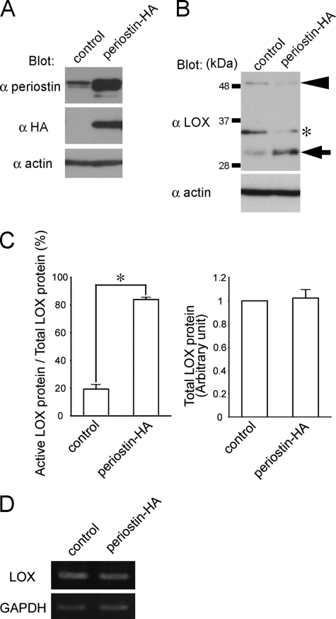
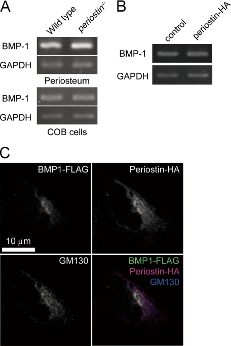
Intra- and intermolecular covalent cross-linking between collagen fibrils, catalyzed by lysyl oxidase (LOX), determines the mechanical properties of connective tissues; however, mechanisms that regulate the collagen cross-linking according to tissue specificity are not well understood. Here we show that periostin, a secretory protein in the dense connective tissues, promotes the activation of LOX. Previous studies showed that periostin null mice exhibit reduced collagen cross-linking in their femurs, periosteum, infarcted myocardium, and tendons. Presently, we showed that active LOX protein, formed by cleavage of its propeptide by bone morphogenetic protein-1 (BMP-1), was decreased in calvarial osteoblast cells derived from periostin null mice. Overexpression of periostin promoted the proteolytic cleavage of the propeptide, which increased the amount of active LOX protein. The results of co-immunoprecipitation and solid phase binding assays revealed that periostin interacted with BMP-1. Furthermore, this interaction probably resulted in enhanced deposition of BMP-1 on the extracellular matrix, suggesting that this enhanced deposition would lead to cleavage of the propeptide of LOX. Thus, we demonstrated that periostin supported BMP-1-mediated proteolytic activation of LOX on the extracellular matrix, which promoted collagen cross-linking.


Figures

FIGURE 1.
FIGURE 1.
Decreased amount of active LOX protein in periostin−/− COB cells. A, expression of LOX family genes in WT and periostin−/− periosteum. Semi-quantitative RT-PCR analysis shows no significant difference in the expression of LOX family genes between WT and periostin−/− periosteum from 12-week-old mice. B, decreased amount of active LOX protein in periostin−/− COB cells. Total cell lysates from WT and periostin−/− COB cells were separated by SDS-PAGE and blotted with anti-LOX and anti-β-actin antibodies. The arrow indicates active LOX, the arrowhead indicates N-glycosylated pro-LOX, and the asterisk indicates the intermediate processed form of LOX. C, quantification of the total and active LOX proteins in WT and periostin−/− COB cells. The independent experiments (n = 4) as shown in B were performed, and the bands corresponding to the total and active LOX proteins were analyzed by densitometric scan, using β-actin for normalization. The asterisks indicate a significant difference (p < 0.05). D, expression of LOX gene in WT and periostin−/− COB cells. Semi-quantitative RT-PCR analysis shows no significant difference in the expression of the LOX gene. GAPDH, glyceraldehyde-3-phosphate dehydrogenase.
FIGURE 2.
FIGURE 2.
Overexpression of periostin-HA increased the amount of active LOX protein in C3H10T1/2 cells. A, expression of periostin-HA in C3H10T1/2 transfectants harboring empty vector (control) or the periostin-HA expression vector (periostin-HA). Total cell lysates were separated with SDS-PAGE and blotted with anti-HA, anti-RD1, and anti-β-actin antibodies. Western blot analysis shows endogenous periostin and overexpression of periostin-HA. B, overexpression of periostin-HA increases the amount of active LOX protein in the C3H10T1/2 transfectants. Total cell lysates were separated by SDS-PAGE and blotted with anti-LOX antibody. The arrow indicates active LOX, the arrowhead indicates N-glycosylated pro-LOX, and the asterisk indicates an intermediate processed form of LOX. C, quantification of the total and active LOX proteins in the C3H10T1/2 transfectants. The independent experiments (n = 4) as shown in B were performed, and the bands corresponding to the total and active LOX proteins were analyzed by densitometric scan, using β-actin for normalization. The asterisk indicates a significant difference (p < 0.0001). D, expression of LOX gene in 10T1/2-control and 10T1/2-periostin-HA. Semi-quantitative RT-PCR analysis shows no significant difference in the expression of LOX gene between the two groups. GAPDH, glyceraldehyde-3-phosphate dehydrogenase.
FIGURE 3.
FIGURE 3.
Co-localization of periostin and BMP-1 in the Golgi. A, expression of BMP-1 gene in periosteum and COB cells from WT and periostin−/− mice. Semi-quantitative RT-PCR analysis shows no significant difference in the expression of the BMP-1 gene in either periosteum or COB cells between the two groups. B, expression of BMP-1 gene in 10T1/2-control and -periostin-HA cells. Semi-quantitative RT-PCR analysis shows no significant difference in the expression of the BMP-1 gene between the two groups. C, co-localization of periostin and BMP-1 in the Golgi. 293 cells were transfected with the expression vectors for periostin-HA and BMP-1-FLAG and then fluorescently stained with anti-HA, anti-FLAG, and anti-GM130 antibodies. GAPDH, glyceraldehyde-3-phosphate dehydrogenase.
FIGURE 4.
FIGURE 4.
Interaction between the fas1 domain of periostin and the metalloproteinase domain of BMP-1. A, schematic view of domain structures of the intact and domain-deletion forms of periostin. These intact and deletion forms were expressed as C-terminal HA-tagged proteins. B, the four tandem repeats of the fas1 domain bind to BMP-1-FLAG. 293 cells were transfected with the expression vectors for periostin-HA or its deletion forms and BMP-1-FLAG. Total cell lysates of the 293 transfectants were subjected to a co-immunoprecipitation (IP) assay with anti-HA antibody-conjugated agarose. Immunoprecipitated complexes were separated by SDS-PAGE and then blotted with anti-HA and anti-FLAG antibodies, respectively. C, schematic view of domain structures of the intact and domain deletion forms of BMP-1. These intact and deletion forms were expressed as C-terminal FLAG-tagged proteins. S, signal sequence; P, prodomain; M, metalloproteinase domain; C1–3, CUB1–3 domains; E, EGF-like domain; Sp, specific region. D, the metalloproteinase domain of BMP-1-FLAG binds to periostin-HA. Co-immunoprecipitation assays between periostin-HA and BMP-1-FLAG or its deletion forms were performed with cell lysates of the 293 transfectants. E, the direct interaction between ΔEMIΔCTR-HA and BMP-1-FLAG in a solid phase. The purified BMP-1-FLAG (open circle) or bovine serum albumin (closed circle) was coated onto wells of the microtiter plate at 5 μg/ml. Binding of ΔEMIΔCTR-HA (0–100 nm) was performed at 4 °C overnight. The bound ΔEMIΔCTR-HA was detected with anti-HA antibody, followed by horseradish peroxidase-conjugated secondary antibody. The error bars represent the means ± S.E.
FIGURE 5.
FIGURE 5.
Periostin increases the deposition of BMP-1 on the fibronectin matrix. A, localization of BMP-1 and fibronectin in the C3H10T1/2 transfectant cell cultures. The C3H10T1/2 transfectants harboring the empty vector (control), the periostin-HA expression vector (periostin-HA), or the periostin(W65A)-HA expression vector (periostin(Trp65Ala)-HA) were grown to 2 days' post-confluence and fluorescently stained with anti-BMP-1 and anti-fibronectin antibodies without prior detergent treatment. The nuclei were stained with TOPRO3. The arrowheads indicate strong fluorescent signals for BMP-1 on the fibronectin matrix. B, overexpression of periostin-HA increases the amount of BMP-1 protein in the total cell lysate and decreases that in the cell culture supernatant (sup.) of the C3H10T1/2 transfectants. The total cell lysates and cell culture supernatants of the C3H10T1/2 transfectants were separated by SDS-PAGE and blotted with anti-BMP-1 and anti-β-actin antibodies, respectively. C, C3H10T1/2 transfectants harboring the empty vector (control), the periostin-HA expression vector (periostin-HA), or the periostin(W65A)-HA expression vector (periostin(Trp65Ala)-HA) were grown to 2 days' post-confluence and quantitatively analyzed for the total amount of LOX-mediated pyridinium cross-linking (n = 5). PYD, pyridinoline; DPD, deoxypyridinoline. The error bars represent the means ± S.E. The asterisk indicates a significant difference (p < 0.0001).

References

    1. Canty E. G., Kadler K. E. (2005) J. Cell Sci. 118, 1341–1353 - PubMed
    1. Banse X., Sims T. J., Bailey A. J. (2002) J. Bone Miner. Res. 17, 1621–1628 - PubMed
    1. Saito M., Fujii K., Mori Y., Marumo K. (2006) Osteoporos. Int. 17, 1514–1523 - PubMed
    1. Seeman E., Delmas P. D. (2006) N. Engl. J. Med. 354, 2250–2261 - PubMed
    1. Açil Y., Vetter U., Brenner R., Müller P. K., Brinckmann J. (1995) J. Am. Acad. Dermatol. 33, 522–524 - PubMed

Publication types