. 2010 Apr 16;285(16):11760-4.
doi: 10.1074/jbc.C109.097220. Epub 2010 Feb 24.

Rho kinase inhibition rescues the endothelial cell cerebral cavernous malformation phenotype

Affiliations

Rho kinase inhibition rescues the endothelial cell cerebral cavernous malformation phenotype

Asya L Borikova et al. J Biol Chem. .

Abstract

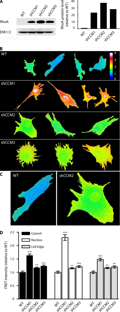
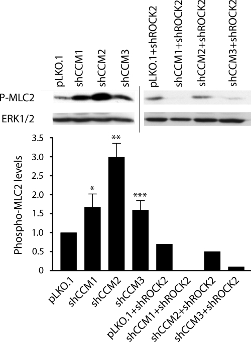
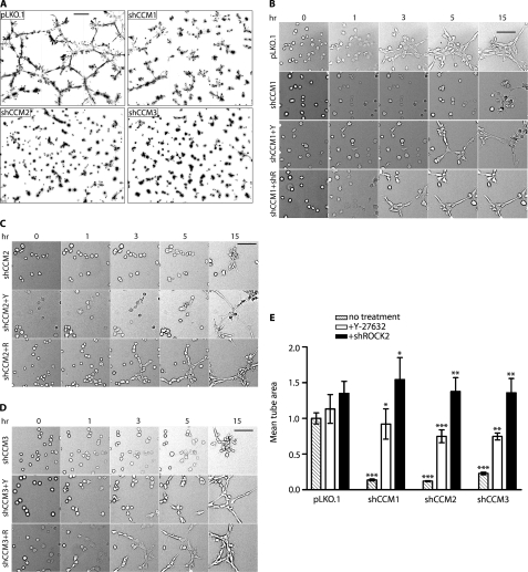
Cerebral cavernous malformations (CCM) are vascular lesions causing seizures and stroke. Mutations causing inactivation of one of three genes, ccm1, -2, or -3, are sufficient to induce vascular endothelial cell defects resulting in CCM. Herein, we show that loss of expression of the CCM1, -2, or -3 proteins causes a marked increase in expression of the GTPase RhoA. Live cell imaging with a RhoA-specific biosensor demonstrates increased RhoA activity with loss of CCM1, -2, or -3, with an especially pronounced RhoA activation in both the cytosol and the nucleus with loss of CCM1 expression. Increased RhoA activation was associated with Rho kinase-dependent phosphorylation of myosin light chain 2. Functionally, loss of CCM1, -2, or -3 inhibited endothelial cell vessel-like tube formation and extracellular matrix invasion, each of which is rescued by chemical inhibition or short hairpin RNA knockdown of Rho kinase. The findings, for the first time, define a signaling network for CCM1, -2, and -3 in CCM pathology, whereby loss of CCM1, -2, or -3 protein expression results in increased RhoA activity, with the activation of Rho kinase responsible for endothelial cell dysregulation. The results define Rho kinase as a therapeutic target to rescue endothelial cells from loss of CCM protein function.


Figures

FIGURE 1.
FIGURE 1.
RhoA abundance and activity are increased in shCCM1, -2, or -3 endothelial cells. A, Western blot showing that the abundance of RhoA in shCCM1, -2, or -3 cells is increased 23-, 37-, and 28-fold relative to WT cells. B, WT, shCCM1, -2, or -3 cells were infected with a FRET-based RhoA biosensor, where activation of RhoA leads to FRET. The measured FRET signal has been pseudocolored, where blue indicates low FRET and low RhoA activity, and red/white indicates high FRET and high RhoA activity. RhoA activity is increased at the cell edge (defined as 1.5-μm width at the edge of the cell), the cytoplasm, and the nucleus of shCCM1, -2, or -3 endothelial cells. C, enlarged view of a representative control and shCCM2 knockdown cell showing active RhoA at the cell edge, cytoplasm, and nucleus. D, bar graph showing the -fold change in FRET intensity for the cytoplasm, nucleus, and cell edge of shCCM1, -2, or -3 relative to WT control cells. Cytoplasm -fold FRET increase values are: shCCM1 cells = 1.62 (p value 4 × 10⁻¹¹), shCCM2 cells = 1.16 (p value 0.001), shCCM3 cells = 1.23 (p value 0.0003). Nuclear -fold FRET increases are: shCCM1 cells = 2.33 (p value 4.8 × 10⁻¹¹), shCCM2 cells = 1.16 (p value 0.002), shCCM3 cells = 1.23 (p value 0.0002). Cell edge -fold FRET increases are: shCCM1 cells = 1.48 (p value 5 × 10⁻⁹), shCCM2 cells = 1.16 (p value 0.002), shCCM3 cells = 1.20 (p value 0.002). Data represent the mean ± S.E. for a minimum of 25 cells in two independent experiments. Error bars, ***, p < 0.001, **, p < 0.02, *, p < 0.05.
FIGURE 2.
FIGURE 2.
Western blots showing an increase in phospho-MLC2 in shCCM1, -2, or -3 endothelial cells, which is lost upon shRNA knockdown of ROCK2. The bar graph shows the densitometric quantitation of band intensity. The abundance of phospho-MLC2 (P-MLC2) in shCCM1, -2, or -3 cells increases 1.7-, 3.0-, and 1.6-fold, respectively, relative to control pLKO.1 cells (the graph represents an average of three independent experiments). Upon shRNA knockdown of ROCK2, this abundance decreases below detectable levels, 50 and 90%, respectively, for shCCM1, -2, or -3 relative to pLKO.1. Error bars, ***, p < 0.001, **, p < 0.02, *, p < 0.05. Line indicates two separate blots.
FIGURE 3.
FIGURE 3.
ROCK inhibitor Y-27632 and ROCK2 shRNA rescue tube formation in shCCM1, -2, or -3 endothelial cells. A, knockdown of CCM1, -2, or -3 results in loss of tube formation. The image is a nine-panel montage of ×10 frames, and the bar represents 300 μm. B, tube formation in shCCM1 cells can be rescued with Y-27632 (+Y) or shROCK2 (+R). Frames are from supplemental Movie 1 taken at 0, 1, 3, 5, and 15 h after the start of treatment with Y-27632. The bar represents 100 μm. The image is a single ×10 field. C, tube formation rescue in shCCM2 cells by shROCK2 or Y-27632. D, tube formation rescue in shCCM3 cells by shROCK2 or Y-27632. E, the bar graph represents the mean tube area of pLKO.1 and shCCM1, -2, and -3 cells, normalized to pLKO.1. The data are the means ± S.E. of six frames from at least three independent experiments. Error bars, ***, p < 0.001, **, p < 0.02, *, p < 0.05.

References

    1. Plummer N. W., Zawistowski J. S., Marchuk D. A. (2005) Curr. Neurol. Neurosci. Rep. 5, 391–396 - PubMed
    1. Laberge-le Couteulx S., Jung H. H., Labauge P., Houtteville J. P., Lescoat C., Cecillon M., Marechal E., Joutel A., Bach J. F., Tournier-Lasserve E. (1999) Nat. Genet. 23, 189–193 - PubMed
    1. Hilder T. L., Malone M. H., Bencharit S., Colicelli J., Haystead T. A., Johnson G. L., Wu C. C. (2007) J. Proteome Res. 6, 4343–4355 - PubMed
    1. Zawistowski J. S., Stalheim L., Uhlik M. T., Abell A. N., Ancrile B. B., Johnson G. L., Marchuk D. A. (2005) Hum. Mol. Genet. 14, 2521–2531 - PubMed
    1. Uhlik M. T., Abell A. N., Johnson N. L., Sun W., Cuevas B. D., Lobel-Rice K. E., Horne E. A., Dell'Acqua M. L., Johnson G. L. (2003) Nat. Cell Biol. 5, 1104–1110 - PubMed

Publication types

MeSH terms

LinkOut - more resources