

The retinal pigment epithelium: something more than a constituent of the blood-retinal barrier--implications for the pathogenesis of diabetic retinopathy
- PMID: 20182540
- PMCID: PMC2825554
- DOI: 10.1155/2010/190724
The retinal pigment epithelium: something more than a constituent of the blood-retinal barrier--implications for the pathogenesis of diabetic retinopathy
Abstract
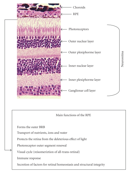
The retinal pigment epithelium (RPE) is an specialized epithelium lying in the interface between the neural retina and the choriocapillaris where it forms the outer blood-retinal barrier (BRB). The main functions of the RPE are the following: (1) transport of nutrients, ions, and water, (2) absorption of light and protection against photooxidation, (3) reisomerization of all-trans-retinal into 11-cis-retinal, which is crucial for the visual cycle, (4) phagocytosis of shed photoreceptor membranes, and (5) secretion of essential factors for the structural integrity of the retina. An overview of these functions will be given. Most of the research on the physiopathology of diabetic retinopathy has been focused on the impairment of the neuroretina and the breakdown of the inner BRB. By contrast, the effects of diabetes on the RPE and in particular on its secretory activity have received less attention. In this regard, new therapeutic strategies addressed to modulating RPE impairment are warranted.
Figures
References
-
- Strauss O. The retinal pigment epithelium in visual function. Physiological Reviews. 2005;85(3):845–881. - PubMed
-
- Steinberg RH. Interactions between the retinal pigment epithelium and the neural retina. Documenta Ophthalmologica. 1985;60(4):327–346. - PubMed
-
- Holtkamp GM, Kijlstra A, Peek R, de Vos AF. Retinal pigment epithelium-immune system interactions: cytokine production and cytokine-induced changes. Progress in Retinal and Eye Research. 2001;20(1):29–48. - PubMed
-
- Congdon NG, Friedman DS, Lietman T. Important causes of visual impairment in the world today. Journal of the American Medical Association. 2003;290(15):2057–2060. - PubMed
-
- Lightman S, Towler HMA. Diabetic retinopathy. Clinical Cornerstone. 2003;5(2):12–21. - PubMed
Publication types
MeSH terms
Substances
LinkOut - more resources
Full Text Sources
Other Literature Sources
Medical
