Review
. 2010:2010:795385.
doi: 10.1155/2010/795385. Epub 2010 Feb 17.

Epidermal growth factor (EGF) treatment on multipotential stromal cells (MSCs). Possible enhancement of therapeutic potential of MSC

Affiliations
Review

Epidermal growth factor (EGF) treatment on multipotential stromal cells (MSCs). Possible enhancement of therapeutic potential of MSC

Kenichi Tamama et al. J Biomed Biotechnol. 2010.

Abstract

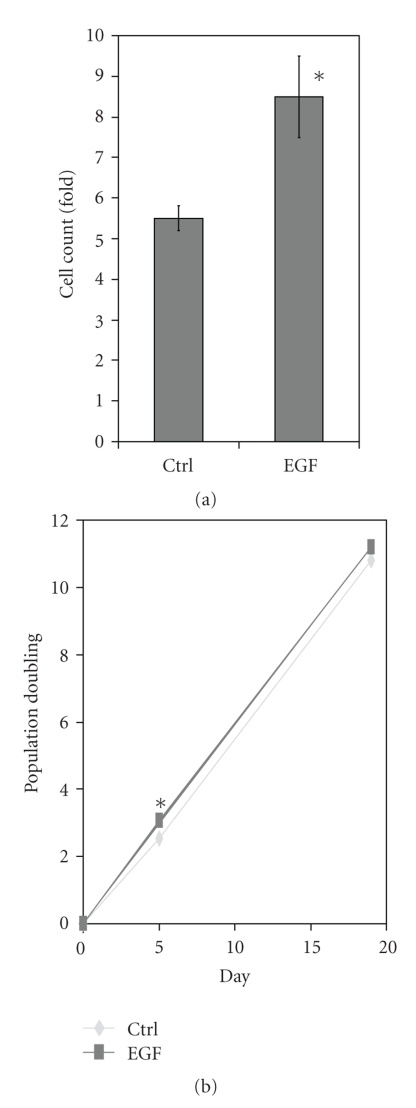
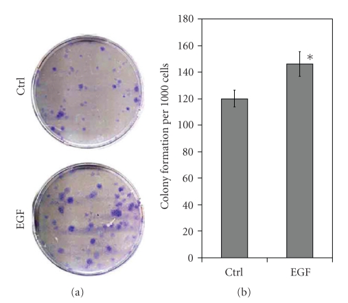
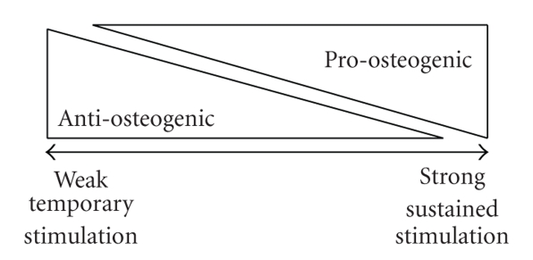
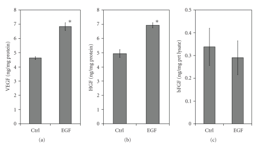
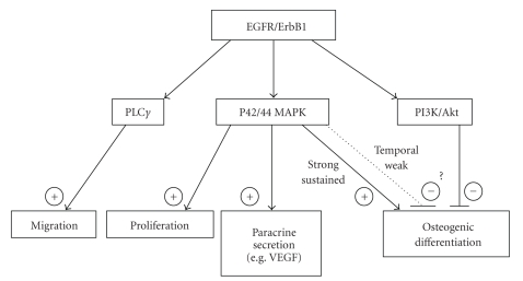
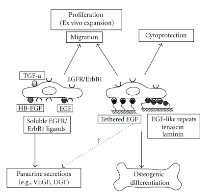
Adult bone marrow multipotential stromal cells (MSCs) hold great promise in regenerative medicine and tissue engineering. However, due to their low numbers upon harvesting, MSCs need to be expanded in vitro without biasing future differentiation for optimal utility. In this concept paper, we focus on the potential use of epidermal growth factor (EGF), prototypal growth factor for enhancing the harvesting and/or differentiation of MSCs. Soluble EGF was shown to augment MSC proliferation while preserving early progenitors within MSC population, and thus did not induce differentiation. However, tethered form of EGF was shown to promote osteogenic differentiation. Soluble EGF was also shown to increase paracrine secretions including VEGF and HGF from MSC. Thus, soluble EGF can be used not only to expand MSC in vitro, but also to enhance paracrine secretion through drug-releasing MSC-encapsulated scaffolds in vivo. Tethered EGF can also be utilized to direct MSC towards osteogenic lineage both in vitro and in vivo.


Figures

Figure 1
Figure 1
Effect of EGF on primary human MSC proliferation (a) and accumulative population doubling (PD)(b). (a) The cell number of primary human MSCs increases about 5.5-fold in the diluent (Ctrl) in culture medium supplemented with 17% FBS in 120 hours (5 days) time period. The addition of EGF (10 nM) gives an extra increase of cell counts to 8.5-fold. Total of 10000 cells was seeded per each well in 12-well plate and the cell count of each well was measured by Coulter Cell Counter Z2 (Beckman Coulter, Inc. Fullerton, CA). Shown are mean ± s.e.m. of three experiments; each performed in triplicate. The differences in proliferation were compared between growth factor and diluent (Ctrl) exposed (*P < .05). (b) Accumulated PD of primary human MSCs in day 5 and 19 in the culture condition same as (a). After cell counting at day 5, equal number of cells (Total of 1000) was seeded per each well in 6-well plate and the cell count of each well was measured at day 19 by Coulter Cell Counter Z2 (Beckman Coulter, Inc. Fullerton, CA). Note that the PD is higher in EGF treated group (2.54 in Ctrl, 3.07 in EGF) for the initial 5 days (*P < .05), but this difference is reduced in the following 14-day period (10.79 in Ctrl, 11.19 in EGF)(No significant difference). Shown are representative data of three independent experiments; each performed in triplicate.
Figure 2
Figure 2
Effects of EGF treatment (10 nM) on primary human MSC colony formation. Five hundred cells were seeded in 10 cm dish within culture medium supplemented with 17% FBS and the number of formed colonies (Diameter ≥1.5 mm) was counted manually in day 14. (a) Representative image of MSC colonies stained with crystal violet. (b) Colony count of MSC. The number of colony is given per 1000 cells seeded initially (*P < .05 to EGF treatment). Shown are representative data of two independent experiments; each performed in triplicate.
Figure 3
Figure 3
Simplified model for the effects of EGFR/ErbB1 signaling on MSC osteogenic differentiation. Weak and temporal stimulation of EGFR/ErbB1 exerts anti-osteogenic effects, whereas strong and sustained stimulation of EGFR/ErbB1 exerts pro-osteogenic effects on MSC.
Figure 4
Figure 4
Effects of EGF treatment (10 nM) on paracrine activities of human primary MSCs. MSCs were cultured in serum-free culture medium with and without EGF (10 nM) for 24 hours. Concentrations of VEGF (a), HGF (b), and bFGF (c) within conditioned media were measured by ELISA and standardized to the total amount of cellular protein contents (*P < .05 to EGF treatment). Shown are average data of three independent experiments; each performed in triplicate.
Figure 5
Figure 5
Simplified diagram of EGFR/ErbB1 signaling pathways in MSC physiology. EGFR/ErbB1 ligands activate PLCγ pathway, p42/44 MAPK pathway, and PI3K/Akt pathways in MSCs [3]. PLCγ pathway plays a pivotal role in motogenic activity, whereas p42/44 MAPK pathway plays a key role in mitogenic activity and paracrine activities of certain factors such as VEGF [42, 43, 66, 67]. Sustained and strong activation of p42/44 MAPK pathway exerts cytoprotective and pro-osteogenic effects [44, 47], whereas PI3K/Akt pathway might exert anti-osteogenic effects [46].
Figure 6
Figure 6
The roles of soluble and tethered EGFR/ErbB1 ligands on MSC physiology. Soluble EGFR/ErbB1 ligands (EGF, HB-EGF, TGF-α) enhance paracrine secretions, proliferation, and migration of MSCs. Tethered EGF or EGF-like repeats within tenascin or laminin augment osteogenic differentiation of MSCs, in addition to cytoprotective, motogenic, and mitogenic effects on MSCs.

Similar articles

Cited by

References

    1. Barry FP, Murphy JM. Mesenchymal stem cells: clinical applications and biological characterization. International Journal of Biochemistry and Cell Biology. 2004;36(4):568–584. - PubMed
    1. Phinney DG, Prockop DJ. Concise review: mesenchymal stem/multipotent stromal cells: the state of transdifferentiation and modes of tissue repair—current views. Stem Cells. 2007;25(11):2896–2902. - PubMed
    1. Tamama K, Fan VH, Griffith LG, Blair HC, Wells A. Epidermal growth factor as a candidate for ex vivo expansion of bone marrow-derived mesenchymal stem cells. Stem Cells. 2006;24(3):686–695. - PubMed
    1. Prockop DJ. Marrow stromal cells as stem cells for nonhematopoietic tissues. Science. 1997;276(5309):71–74. - PubMed
    1. Pittenger MF, Mackay AM, Beck SC, et al. Multilineage potential of adult human mesenchymal stem cells. Science. 1999;284(5411):143–147. - PubMed

Publication types

LinkOut - more resources