

Examination of the role of magnetic resonance imaging in multiple sclerosis: A problem-orientated approach
- PMID: 20182573
- PMCID: PMC2824953
- DOI: 10.4103/0972-2327.58284
Examination of the role of magnetic resonance imaging in multiple sclerosis: A problem-orientated approach
Abstract
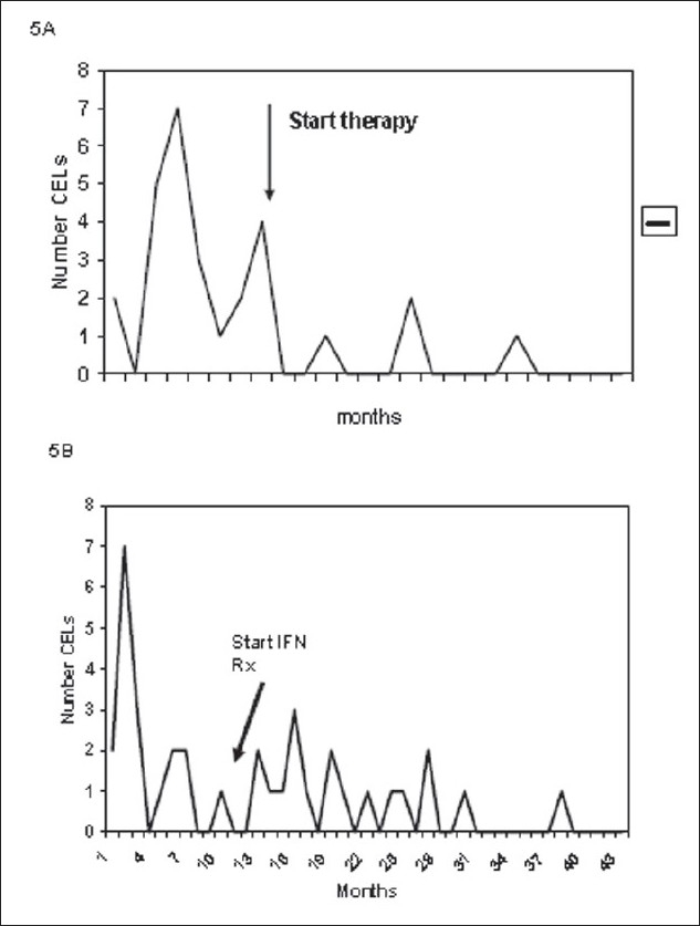
Magnetic Resonance Imaging (MRI) has brought in several benefits to the study of Multiple Sclerosis (MS). It provides accurate measurement of disease activity, facilitates precise diagnosis, and aid in the assessment of newer therapies. The imaging guidelines for MS are broadly divided in to approaches for imaging patients with suspected MS or clinically isolated syndromes (CIS) or for monitoring patients with established MS. In this review, the technical aspects of MR imaging for MS are briefly discussed. The imaging process need to capture the twin aspects of acute MS viz. the autoimmune acute inflammatory process and the neurodegenerative process. Gadolinium enhanced MRI can identify acute inflammatory lesions precisely. The commonly applied MRI marker of disease progression is brain atrophy. Whole brain magnetization Transfer Ratio (MTR) and Magnetic Resonance Spectroscopy (MRS) are two other techniques use to monitor disease progression. A variety of imaging techniques such as Double Inversion Recovery (DIR), Spoiled Gradient Recalled (SPGR) acquisition, and Fluid Attenuated Inversion Recovery (FLAIR) have been utilized to study the cortical changes in MS. MRI is now extensively used in the Phase I, II and III clinical trials of new therapies. As the technical aspects of MRI advance rapidly, and higher field strengths become available, it is hoped that the impact of MRI on our understanding of MS will be even more profound in the next decade.
Keywords: Magnetic resonance imaging; multiple sclerosis; problem-oriented clinical approach.
Conflict of interest statement
Figures
Similar articles
-
[Magnetic resonance imaging in multiple sclerosis].Pathol Biol (Paris). 2000 Mar;48(2):151-61. Pathol Biol (Paris). 2000. PMID: 10815291 Review. French.
-
The role of magnetic resonance techniques in understanding and managing multiple sclerosis.Brain. 1998 Jan;121 ( Pt 1):3-24. doi: 10.1093/brain/121.1.3. Brain. 1998. PMID: 9549485 Review.
-
Advanced MRI Methods for Diagnosis and Monitoring of Multiple Sclerosis (MS).J Magn Reson Imaging. 2025 May 27. doi: 10.1002/jmri.29817. Online ahead of print. J Magn Reson Imaging. 2025. PMID: 40424444 Review.
-
Detection of Cortical and Deep Gray Matter Lesions in Multiple Sclerosis Using DIR and FLAIR at 3T.J Neuroimaging. 2021 Mar;31(2):408-414. doi: 10.1111/jon.12822. Epub 2020 Dec 22. J Neuroimaging. 2021. PMID: 33351983
-
Magnetic resonance imaging in multiple sclerosis.Rev Neurol Dis. 2005 Summer;2(3):109-16. Rev Neurol Dis. 2005. PMID: 16400309 Review.
Cited by
-
Therapies for multiple sclerosis: considerations in the pediatric patient.Nat Rev Neurol. 2011 Feb;7(2):109-22. doi: 10.1038/nrneurol.2010.198. Epub 2011 Jan 11. Nat Rev Neurol. 2011. PMID: 21224883 Review.
-
Current Methods of Magnetic Resonance for Noninvasive Assessment of Molecular Aspects of Pathoetiology in Multiple Sclerosis.Int J Mol Sci. 2020 Aug 25;21(17):6117. doi: 10.3390/ijms21176117. Int J Mol Sci. 2020. PMID: 32854318 Free PMC article. Review.
-
Gradient echo plural contrast imaging--signal model and derived contrasts: T2*, T1, phase, SWI, T1f, FST2*and T2*-SWI.Neuroimage. 2012 Apr 2;60(2):1073-82. doi: 10.1016/j.neuroimage.2012.01.108. Epub 2012 Jan 28. Neuroimage. 2012. PMID: 22305993 Free PMC article.
-
Magnetic Resonance Imaging in Multiple Sclerosis.Cold Spring Harb Perspect Med. 2018 May 1;8(5):a028969. doi: 10.1101/cshperspect.a028969. Cold Spring Harb Perspect Med. 2018. PMID: 29358319 Free PMC article. Review.
-
Lifestyle management and brain MRI metrics in female Australian adults living with multiple sclerosis: a feasibility and acceptability study.Pilot Feasibility Stud. 2024 May 2;10(1):71. doi: 10.1186/s40814-024-01495-3. Pilot Feasibility Stud. 2024. PMID: 38698454 Free PMC article.
References
-
- Harris JO, Frank JA, Patronas N, McFarlin DE, McFarland HF. Serial gadolinium-enhanced magnetic resonance imaging scans in patients with early, relapsing-remitting multiple sclerosis: Implications for clinical trials and natural history. Ann Neurol. 1991;29:548–55. - PubMed
-
- Miller DH, Barkhof F, Nauta JJ. Gadolinium enhancement increases the sensitivity of MRI in detecting disease activity in multiple sclerosis. Brain. 1993;116:1077–94. - PubMed
-
- Katz D, Taubenberger JK, Cannella B, McFarlin DE, Raine CS, McFarland HF. Correlation between magnetic resonance imaging findings and lesion development in chronic, active multiple sclerosis. Ann Neurol. 1993;34:661–9. - PubMed
-
- Frank JA, Stone LA, Smith ME, Albert PS, Maloni H, McFarland HF. Serial contrast-enhanced magnetic resonance imaging in patients with early relapsing-remitting multiple sclerosis: Implications for treatment trials. Ann Neurol. 1994;36:S86–90. - PubMed
LinkOut - more resources
Full Text Sources
Research Materials
