

Molecular basis of the core structure of tight junctions
- PMID: 20182608
- PMCID: PMC2827901
- DOI: 10.1101/cshperspect.a002907
Molecular basis of the core structure of tight junctions
Abstract
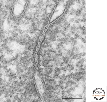
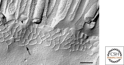
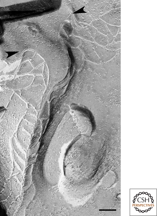
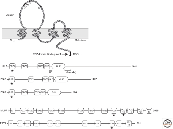
The morphological feature of tight junctions (TJs) fits well with their functions. The core of TJs is a fibril-like proteinaceous structure within the lipid bilayer, the so-called TJ strands. TJ strands in apposing plasma membranes associate with each other to eliminate the intercellular space. A network of paired TJ strands generates a continuous belt that circumscribes each cell to establish the diffusion barrier to the solutes in the paracellular pathway throughout the cellular sheet. Identification and characterization of TJ-associated proteins during the last two decades has unveiled the nature of TJ strands and how they are spatially organized. The interplay between integral membrane proteins, claudins, and cytoplasmic plaque proteins, ZO-1/ZO-2, is critical for TJ formation and function.
Figures
References
-
- Anderson JM, Cereijido M 2001. Evolution of ideas on the tight junction. In Tight junctions, 2nd ed. (ed. Cereijido M, Anderson J), pp. 1–18 CRC Press, Boca Raton
-
- Aono S, Hirai Y 2008. Phosphorylation of claudin-4 is required for tight junction formation in a human keratinocyte cell line. Exp Cell Res 314:3326–3339 - PubMed
-
- Balda MS, Anderson JM 1993. Two classes of tight junctions are revealed by ZO-1 isoforms. Am J Physiol 264:918–924 - PubMed
Publication types
MeSH terms
Substances
LinkOut - more resources
Full Text Sources
Other Literature Sources