Review
. 2010 Feb;2(2):a000869.
doi: 10.1101/cshperspect.a000869.

Signaling gradients during paraxial mesoderm development

Affiliations
Review

Signaling gradients during paraxial mesoderm development

Alexander Aulehla et al. Cold Spring Harb Perspect Biol. 2010 Feb.

Abstract

The sequential formation of somites along the anterior-posterior axis is under control of multiple signaling gradients involving the Wnt, FGF, and retinoic acid (RA) pathways. These pathways show graded distribution of signaling activity within the paraxial mesoderm of vertebrate embryos. Although Wnt and FGF signaling show highest activity in the posterior, unsegmented paraxial mesoderm (presomitic mesoderm [PSM]), RA signaling establishes a countergradient with the highest activity in the somites. The generation of these graded activities relies both on classical source-sink mechanisms (for RA signaling) and on an RNA decay mechanism (for FGF signaling). Numerous studies reveal the tight interconnection among Wnt, FGF, and RA signaling in controlling paraxial mesoderm differentiation and in defining the somite-forming unit. In particular, the relationship to a molecular oscillator acting in somite precursors in the PSM-called the segmentation clock-has been recently addressed. These studies indicate that high levels of Wnt and FGF signaling are required for the segmentation clock activity. Furthermore, we discuss how these signaling gradients act in a dose-dependent manner in the progenitors of the paraxial mesoderm, partly by regulating cell movements during gastrulation. Finally, links between the process of axial specification of vertebral segments and Hox gene expression are discussed.


Figures

Figure 1.
Figure 1.
Scheme of signaling gradient system in the presomitic mesoderm (PSM) of mouse embryos. If not stated otherwise, illustrations reflect mRNA expression patterns in the PSM of mouse embryos. Additional sites of expression (e.g., somites, neural tube) are not shown. For Akt and Erk, the distribution of the activated, phosphorylated protein is shown (p-Akt, p-Erk). For Erk, these data derive from chicken (c) and zebrafish (z) embryos. For Axin2, Dusp6, and Dusp4, the graded expression is periodically down-regulated in the posterior PSM, as indicated by the oscillation symbol (∼).
Figure 2.
Figure 2.
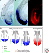
An RNA decay mechanism establishes the Fgf8 gradient within the PSM. (A) In situ hybridization (ISH) for Fgf8 pre-mRNA using an intronic probe in a mouse embryo indicates that de novo transcription is localized to the tail bud region. (S1) last formed somite, (PSM) presomitic mesoderm. (B) ISH for mature Fgf8 mRNA shows a graded distribution within the PSM. (C) Immunofluorescent detection of Fgf8 shows a posterior-anterior protein gradient. (D) Scheme of gradient formation using an RNA decay mechanism. In the temporal series ac, a constant group of cells (orange square) and its Fgf8 RNA expression characteristics are shown. In a, this group of cells is located in the posterior-most PSM and tail bud and hence shows de novo Fgf8 transcription. For simplicity, only de novo synthesized RNA is shown in a. At a later time point (b), the same group of cells is now located in the posterior one-third of PSM because of the posterior addition of cells during axis elongation. Fgf8 transcription in the PSM ceased, and as a consequence, Fgf8 mRNA decayed in comparison to the levels in the tail bud. At an even later time point (c), this group of cells is located in the middle PSM, and again Fgf8 mRNA levels are further decreased. In consequence, Fgf8 mRNA levels are graded in the PSM. This mRNA gradient is translated into a posterior-anterior Fgf8 ligand gradient (d).
Figure 3.
Figure 3.
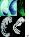
A β-catenin protein gradient controls PSM differentiation and somite formation. (A) ISH for Wnt3a mRNA in 9.5 days postcoitum (dpc) mouse embryo shows site of localized ligand production in the posterior PSM/tail bud. (B) ISH for β-catenin mRNA shows ubiquitous expression in the PSM of mouse embryos. (C) In contrast, β-catenin immunofluorescent detection (green) reveals a posterior-anterior nuclear protein gradient within the PSM. DAPI staining is shown in blue. (D) Scanning electron microscopy image of a control 9.0 dpc mouse embryo showing the periodic arrangement of formed somites. (E) Scanning electron microscopy image of mutant embryo in which a stabilized isoform of β-catenin has been conditionally expressed in mesodermal cells. Note that disruption of the β-catenin protein gradient leads to an expanded, undifferentiated PSM and an absence of somite formation. In D and E, the ectoderm was removed, which allows a direct view of the mesodermal layer.
Figure 4.
Figure 4.
Model for segment determination using opposing gradients to create a bistability domain. Two steady states are proposed to coexist in the PSM. In the posterior part, cells adopt a fibroblast growth factor (FGF)/Wnt-dominated state (purple), whereas in the anterior PSM the opposing retinoic acid (RA) state dominates (green). Within a window of bistability (dashed rectangle), cells can be triggered to abruptly switch between either of the two steady states. This trigger is proposed to be provided by the periodic signal delivered by the segmentation clock. As a result, a cohort of cells in the bistability domain will be exposed simultaneously to higher levels of RA signaling and, thereby, the segment-forming unit becomes defined (orange). Owing to the posterior extension of the axis and the decay of the FGF and Wnt mRNA and ligands in the PSM, the bistability window constantly moves posteriorly. The next cohort of cells to be simultaneously determined is shown in blue. Note that the bistability window is proposed to be larger than the segment-forming unit, because it comprises also the segment that was defined during the previous cycle and therefore had already responded to the clock signal. In this model, the posterior edge of the bistability window (bifurcation point) corresponds to the determination front.
Figure 5.
Figure 5.
Model of temporal gradients in paraxial mesoderm precursors. The posterior region of three mouse embryos at day 9.5 dpc, 10.5 dpc, and 11.0 dpc are shown schematically and the zone of paraxial progenitors (long-term progenitors [LTP]) is highlighted (black box). Within this LTP, Wnt and FGF signaling might increase over time (plotted in orange), for instance, because of progressive accumulation of the ligands. Also, the exposure time of LTP to Wnt and FGF signals might increase over time (plotted in green). In this model, these gradients are proposed to control cell movements during gastrulation (e.g., the timing of how cells exit the primitive streak) and hence control replenishment of the PSM. Together with Hox genes and possibly via Cdx genes, these potential gradients could contribute to axial specification of vertebrae identity. This results in vertebrae with unique features along the AP axis, as shown in a mouse embryo at day 14.5 dpc.

Similar articles

Cited by

References

    1. Arnold SJ, Stappert J, Bauer A, Kispert A, Herrmann BG, Kemler R 2000. Brachyury is a target gene of the Wnt/β-catenin signaling pathway. Mech Dev 91:249–258 - PubMed
    1. Aulehla A, Herrmann BG 2004. Segmentation in vertebrates: Clock and gradient finally joined. Genes Dev 18:2060–2067 - PubMed
    1. Aulehla A, Wehrle C, Brand-Saberi B, Kemler R, Gossler A, Kanzler B, Herrmann BG 2003. Wnt3a plays a major role in the segmentation clock controlling somitogenesis. Dev Cell 4:395–406 - PubMed
    1. Aulehla A, Wiegraebe W, Baubet V, Wahl MB, Deng C, Taketo M, Lewandoski M, Pourquié O 2008. A β-catenin gradient links the clock and wavefront systems in mouse embryo segmentation. Nat Cell Biol 10:186–193 - PMC - PubMed
    1. Barrallo-Gimeno A, Nieto MA 2005. The Snail genes as inducers of cell movement and survival: Implications in development and cancer. Development 132:3151–3161 - PubMed

Publication types

LinkOut - more resources